Introduction
Pediatric cataracts are a leading cause of treatable childhood blindness. If left unaddressed, cataracts can have substantial social, economic, and emotional consequences for the affected child, family, and broader community. Effective management remains challenging due to the critical need for early identification, diagnosis, and intervention to prevent irreversible amblyopia.[1]
Routine screening and parental awareness of early signs, such as leukocoria and strabismus, facilitate timely diagnosis and treatment. Favorable visual outcomes depend on comprehensive preoperative assessment, accurate intraocular lens (IOL) power calculation, meticulous surgical technique, and coordinated postoperative care, including visual rehabilitation. Optimal management requires an interprofessional approach involving pediatrics, anesthesiology, ophthalmology, and optometry.[2][3][4]
Pediatric cataracts are a major cause of preventable childhood blindness worldwide, particularly in developing countries, where delayed diagnosis often leads to advanced presentations such as nystagmus, poor fixation, and dense cataracts. Early intervention significantly improves visual outcomes and enhances the affected child’s personal and social development, while also alleviating the socioeconomic burden on families. Globally, pediatric cataracts account for approximately 5% to 20% of childhood blindness and severe visual impairment, with an estimated incidence of 1.8 to 3.6 per 10,000 children per year and a prevalence ranging from 1 to 15 per 10,000 children.[5]
Population-based data show similar trends in high-income settings. Holmes et al reported a prevalence of 3 to 4 visually significant cataracts per 10,000 live births in the United States.[6] A study by Rahi et al in the United Kingdom found a prevalence of 3.18 per 10,000 live births, while Nile et al reported an incidence of approximately 5 per 10,000 births in China. Despite regional differences in detection and reporting, consistent findings across studies indicate no significant laterality or sex predilection.[7]
Hereditary congenital cataracts have a prevalence between 8.3% and 25%, with about 75% of cases following an autosomal dominant inheritance pattern. Pathogenic variants in crystallin proteins, which are essential for maintaining lens transparency and refractive function, have been associated with several cataract subtypes, including nuclear, lamellar, zonular, and posterior polar cataracts. Nonsyndromic inherited cataracts frequently involve genetic alterations in crystallin or connexin genes. PITX3 mutations are specifically linked to posterior polar cataracts, while PAX6 alterations are associated with anterior polar cataracts.[8]
Syndromic cataracts are linked to specific genetic defects, including galactosidase-α in Fabry disease, galactose-1-phosphate uridyltransferase (GALT) in galactosemia, OCRL in Lowe (oculocerebrorenal) syndrome, and NHS in Nance–Horan syndrome, a cataract–dental disorder (see Image. Congenital Cataracts and Abnormal Galactose Metabolism). Maternal and congenital infections, particularly Toxoplasma gondii, rubella, cytomegalovirus, herpes simplex virus, and Treponema pallidum (syphilis)—collectively known as TORCH pathogens—are also major contributors to pediatric cataracts. B Mahalakshmi et al reported a high prevalence of TORCH infections in the Indian subcontinent, with 20% of cases testing seropositive. Ocular trauma is another significant cause, accounting for 12% to 46% of pediatric cataract cases.[9]
Concerns exist regarding the higher incidence of complications, such as glaucoma, uveitis, dense posterior capsule opacification (PCO), and increased secondary interventions following primary IOL implantation in children younger than 2 years.[10] However, primary IOL implantation in these young individuals has been shown to be safe, with excellent long-term outcomes compared to aphakia and secondary IOL implantation after age 2. The myopic shift is generally well-controlled, visual acuity outcomes are favorable, and the incidence of complications, such as glaucoma, uveitis, membrane formation, synechiae, and the need for secondary interventions, is lower than previously reported. Special care is necessary for children younger than 6 months due to the high risk of adverse events in smaller eyes.[11]
The process of emmetropisation in children is typically complete by age 12, with axial length increasing from approximately 16.5 mm at birth to 23 mm by age 13. This growth occurs in 3 phases: the rapid phase (0.46 mm/month from birth to 6 months), the infantile phase (0.15 mm/month from 6 to 18 months), and the juvenile phase (from 18 months to 12 years).
Corneal curvature also changes significantly, with mean keratometry readings decreasing from approximately 51.2 D at birth to 43.5 D in adulthood. Consequently, the power of the IOL implanted in children must be adjusted to account for both axial elongation and the accompanying myopic shift. This adjustment requires the use of customized IOL power calculation formulas suited to growing pediatric eyes.
Sharp-edged IOLs are now widely preferred due to their association with lower rates of visual axis opacification (VAO), resulting in fewer neodymium-doped yttrium aluminum garnet (Nd:YAG) laser capsulotomies compared to round-edged lenses (1/371 vs 4/371).
Prompt management of pediatric cataracts is essential for optimal visual development. Most children with congenital or developmental cataracts will require surgical intervention. The degree of visual impairment may be initially assessed using the red reflex during distant direct ophthalmoscopy (see Image. Red Reflex). For visually significant cataracts, bilateral cases should be treated between 6 and 8 weeks of age, while unilateral cases require earlier intervention, typically between 4 and 6 weeks.[12]
Etiology
Register For Free And Read The Full Article
 Search engine and full access to all medical articles
- 10 free questions in your specialty
- Free CME/CE Activities
- Free daily question in your email
- Save favorite articles to your dashboard
- Emails offering discounts
Learn more about a Subscription to StatPearls Point-of-Care
Etiology
The etiologies of pediatric cataracts are diverse, ranging from conditions of unknown origin to those associated with genetic or systemic disorders. Cataracts may be unilateral or bilateral, with most unilateral cases and a proportion of bilateral cases classified as idiopathic.
Causes of congenital cataracts include the following:
- Intrauterine infections: T. gondii, rubella, cytomegalovirus, herpes simplex virus, syphilis (TORCHES) [13]
- Drug-induced: Corticosteroids (especially prednisolone), ivacaftor (a potentiator of the cystic fibrosis transmembrane conductance regulator, or CFTR, used to treat cystic fibrosis), and topotecan (a topoisomerase I inhibitor) [14]
- Metabolic disorders: Galactosemia, galactokinase deficiency, hypocalcemia, and hypoglycemia [15]
- Trauma, whether accidental or nonaccidental
- Radiation exposure: Laser photocoagulation
- Association with other ocular conditions: Microphthalmia, microcornea, aniridia, persistent hyperplastic primary vitreous (PHPV), Peter anomaly, corneal guttae, coloboma
- Inherited without systemic abnormalities: Autosomal dominant (most common), autosomal recessive (more common in consanguineous families), and X-linked [16]
- Association with other systemic conditions:
- Chromosomal abnormalities: Trisomies 13, 18, and 21; Turner and Cri-du-chat syndromes
- Cerebro-oculo-facio-skeletal syndrome
- Mitochondrial disorders: Complex I deficiency
- Renal disease: Lowe syndrome
- Skeletal disorders: Smith-Lemli-Opitz, Conradi, and Weill-Marchesani syndromes
- Digital anomalies, such as syndactyly or polydactyly: Bardet-Biedl and Rubinstein-Taybi syndromes
- Central nervous system abnormalities: Zellweger and Meckel-Gruber syndromes
- Cardiac disease: Hypertrophic cardiomyopathy
- Dermatologic disorders: Cockayne and Rothmund-Thomson syndromes, atopic dermatitis, incontinentia pigmenti, progeria, ichthyosis, and ectodermal dysplasia
- Dental anomalies: Nance-Horan and Lenz syndromes
- Idiopathic
Bilateral congenital cataracts may arise in the following settings:
- Idiopathic cases (~60%)
- Hereditary transmission (~30%): Autosomal dominant (~75%; most common), autosomal recessive, and X-linked
- Intrauterine infections (TORCHES)
- Multisystem genetic disorders
- Inborn errors of metabolism
- Endocrinopathies
- Trauma
- Uveitic cataract
- Ocular abnormalities
- Genetic conditions: Down and Turner syndromes, myotonic dystrophy
- Metabolic disorders: Diabetes mellitus (vacuolar cataracts), hypoglycemia, hypocalcemia, galactosemia (oil droplet cataracts), Fabry disease (spoke-like cataracts), Zellweger syndrome, hypoparathyroidism (multicolored flecks), and Lowe syndrome (thin disciform cataracts)
- Drug reactions: Steroids, miotics, chlorpromazine, amiodarone
Pediatric cataracts exhibit a wide range of etiologies and clinical manifestations. A thorough etiologic evaluation is critical since the specific cause dictates clinical management and long-term treatment planning. Recognizing laterality and identifying potential systemic associations are critical for delivering comprehensive care and optimizing visual outcomes.
Epidemiology
Various epidemiological studies have been conducted to evaluate the causes of childhood blindness. Untreated pediatric cataracts account for approximately 7.4% to 15.3% of cases. The incidence and prevalence of pediatric cataracts vary by region, with lower rates in high-income countries and higher rates in low-income settings. Incidence ranges from 1.8 to 3.6 per 10,000 children annually, while prevalence ranges from 0.63 to 13.6 per 10,000 in low-income countries such as Bangladesh, Pakistan, and India, compared to 0.42 to 2.05 per 10,000 in high-income countries such as the United States and the United Kingdom.[17] No significant differences in prevalence have been observed based on sex or laterality.[18]
Pediatric cataracts are a chief cause of treatable childhood blindness globally, with higher incidence rates reported in developing countries. If not promptly treated, the condition can result in severe visual impairment, affecting the child's quality of life and placing socioeconomic strain on the family and broader community. Pediatric cataracts account for an estimated 5% to 20% of childhood blindness and severe visual impairment worldwide. The global incidence ranges from 1.8 to 3.6 per 10,000 children annually, and prevalence is estimated at 1 to 15 per 10,000. Country-specific studies show varied prevalence rates. In the United States, the prevalence is estimated at 3 to 4 visually significant cataracts per 10,000 live births.[19]
The prevalence of pediatric cataracts in the United Kingdom is approximately 3.18 per 10,000 live births. In China, the incidence is around 5 per 10,000 births. Although geographic variations in prevalence exist, significant differences in laterality (ie, whether 1 or both eyes are affected) or sex are not commonly reported.
Hereditary cataracts account for approximately 30% of all pediatric cases, with about 75% following an autosomal dominant inheritance pattern and the remainder inherited in an autosomal recessive or X-linked fashion. Specific genetic conditions associated with pediatric cataracts include Down and Turner syndromes, as well as myotonic dystrophy. Several metabolic disorders also contribute to cataract development, including diabetes mellitus, hypoglycemia, hypocalcemia, galactosemia, Fabry disease, Zellweger syndrome, hypoparathyroidism, and Lowe syndrome.[20]
Certain medications, such as steroids, miotics, chlorpromazine, and amiodarone, can induce cataracts in children. Trauma is another major cause, accounting for 12% to 46% of pediatric cases. Maternal and congenital infections, particularly those from the TORCHES group, are also notable contributors. A study conducted in the Indian subcontinent reported a high incidence of TORCH infections, with 20% of cases testing seropositive.
Pediatric cataracts represent a critical public health concern that requires timely diagnosis and intervention to prevent long-term visual impairment. A thorough understanding of the epidemiology, including genetic, metabolic, drug-induced, traumatic, and infectious cases, is essential for effective management and treatment planning.[21]
Pathophysiology
Pediatric cataracts have diverse causes, including heritable genetic defects, fetal or childhood developmental insults, and associated systemic syndromes. Autosomal dominant inheritance is the most common pattern seen in hereditary cataracts. A genetic screening study conducted in Australia identified as many as 51 genes and loci associated with the condition.[22] Mutations in genes encoding transcription factors, such as PAX6, FOXE3, MAF (C-MAF), PITX3, MIP, and CRYAA, are frequently observed. Mutations in genes encoding crystallin and connexin are also common.[23][24]
Pediatric cataracts cloud the eye's natural lens and impair vision. The multifactorial pathophysiology involves genetic, metabolic, infectious, and environmental factors that disrupt the normal transparency and function of the lens.
Genetic mutations contribute not only to the presence of pediatric cataracts but also to their phenotypic variability and severity. The affected genes encode proteins essential for lens structure, development, and transparency. Crystallins, for instance, are the primary structural proteins of the lens, maintaining its optical clarity and refractive index. Alterations in crystallin genes lead to protein misfolding or aggregation, resulting in distinct cataract morphologies such as nuclear, lamellar, zonular, and posterior polar types. Connexins form gap junctions that facilitate intercellular communication and metabolic balance within the lens. The disruption of these structures impairs homeostasis and contributes to lens opacification.
Transcription factors such as PITX3 and PAX6 orchestrate lens morphogenesis and differentiation. Mutations in PITX3 have been linked to posterior polar cataracts, while PAX6 alterations are more commonly associated with anterior polar variants. These gene-specific effects highlight the complex molecular basis of pediatric cataracts and underscore the need for genotype-phenotype correlation in guiding diagnosis and management.
Pediatric cataracts may also arise from underlying metabolic disorders. Diabetes mellitus causes elevated blood glucose levels, leading to osmotic imbalances in the lens that result in swelling and vacuole formation, ultimately contributing to cataractogenesis. In galactosemia, the accumulation of galactitol from impaired galactose metabolism induces osmotic stress and leads to the formation of oil droplet cataracts. Hypocalcemia, characterized by low calcium levels, disrupts lens cell function and protein stability, promoting cataract formation. Fabry disease, a lysosomal storage disorder, results in glycosphingolipid accumulation and is associated with spoke-like lens opacities.[25]
Maternal and congenital infections, particularly TORCH infections, can lead to congenital cataracts in newborns. These infections induce intrauterine inflammation and damage the developing lens, resulting in opacification.[26]
Drug-induced cataracts may occur following prolonged exposure to specific medications. Corticosteroids, for instance, alter lens metabolism and protein structure, contributing to posterior subcapsular cataracts. Other drugs, such as miotics, chlorpromazine, and amiodarone, can induce lens opacities through oxidative stress or disruption of lens cell function. Trauma to the eye, whether accidental or intentional, can also result in cataract formation. Mechanical injury may disrupt the lens capsule, allowing fluid ingress and protein denaturation that ultimately leads to lens opacification.[27]
Environmental and nutritional factors play significant roles in the development of pediatric cataracts. Prolonged exposure to ultraviolet radiation and other environmental stressors can heighten oxidative stress within the lens. This increased oxidative burden damages lens proteins and lipids, ultimately leading to cataract formation. Additionally, deficiencies in essential nutrients, particularly antioxidants such as vitamins C and E, can compromise lens health. Such deficiencies impair the lens’s ability to counteract oxidative damage and contribute to cataract progression.[28]
The mechanisms underlying lens opacification are multifaceted and involve several biological processes. Protein aggregation, often resulting from genetic mutations or metabolic imbalances, leads to the unfolding and clumping of crystallin proteins, which scatter light and impair lens transparency. Osmotic imbalances, as seen in conditions such as diabetes and galactosemia, cause the accumulation of osmotic agents within the lens, triggering cellular swelling, membrane rupture, and protein leakage. Disruption of calcium homeostasis further contributes to lens opacity by altering cell signaling and destabilizing lens proteins.[29]
Inflammatory responses induced by infections or trauma can damage lens cells and proteins, further promoting cataract formation. Given the complex pathophysiology of pediatric cataracts, which encompasses genetic mutations, metabolic disturbances, infections, drug toxicity, trauma, and environmental influences, a comprehensive understanding of these mechanisms is essential for developing effective preventive and therapeutic strategies to preserve vision in affected children.[30]
Histopathology
The histopathology of pediatric cataracts involves the microscopic examination of lens tissue to characterize the structural changes that lead to opacification. Different cataract types exhibit distinct histopathological features, offering insight into their underlying mechanisms and informing potential treatment approaches.
Pediatric cataracts commonly display disorganized and fragmented lens fibers. In healthy lenses, fibers are tightly packed and transparent. However, in cataractous lenses, the fibers may appear swollen, misaligned, or vacuolated. Aggregates of denatured or misfolded proteins, particularly crystallins, are often observed within lens fibers. These aggregates scatter light and contribute to lens opacity. Fluid-filled vacuoles are frequently seen in pediatric cataracts, especially those associated with metabolic disorders. These vacuoles arise from osmotic imbalance and lens cell damage.[31]
Genetic cataracts manifest in various forms, each with characteristic histological features. Nuclear cataracts exhibit central lens opacity with dense protein aggregation and increased compaction of lens fibers in the nucleus. Lamellar cataracts present as disk-shaped opacities involving specific layers of the lens. Histologically, these layers contain swollen and disrupted fibers with embedded protein aggregates. Posterior polar cataracts are localized to the posterior aspect of the lens and involve abnormalities of the posterior capsule and adjacent lens fibers. Histologic findings include capsular fibrosis and aberrant cellular proliferation, both of which contribute to the observed opacification.
Metabolic cataracts encompass a spectrum of conditions associated with metabolic disturbances, each exhibiting distinctive pathological features. Cataracts related to galactosemia are characterized by oil droplet formations within the lens, accompanied by vacuolization, osmotic swelling of lens fibers, and deposits of galactitol. Diabetic cataracts display hallmark features such as vacuolar changes, fiber swelling, and sorbitol accumulation. The osmotic effect induced by intracellular sorbitol promotes water influx into the lens, leading to opacification. Cataracts associated with Fabry disease often present with spoke-like opacities, lipid accumulation, and abnormal glycosphingolipid deposits within lens fibers. These distinct histological findings underscore the complex relationship between metabolic abnormalities and lens pathology in metabolic cataracts.[32]
Congenital infections, particularly those caused by TORCH pathogens, leave characteristic histopathological imprints on the lens. Microscopic examination typically reveals inflammation, fibrosis, and cellular infiltration within lens tissue. Thickening of the lens capsule may also be observed, along with viral or parasitic inclusion bodies, further highlighting the detrimental effects of congenital infections on lens development and integrity.
Traumatic cataracts arise from physical injury to the eye and elicit various pathological changes within the lens. Capsule rupture, subsequent fibrosis, and abnormal cell proliferation are common posttraumatic findings. Disruption of the lens capsule’s integrity often results in disorganized lens fibers and the development of opacities. In some cases, calcium deposition within the lens further contributes to opacity and increases lens rigidity. These pathological alterations underscore the diverse structural and functional consequences of trauma in the development of traumatic cataracts.[33]
Certain medications can also induce cataract formation. Steroid-induced cataracts typically manifest as posterior subcapsular opacities, with histopathological findings of swollen and vacuolated lens epithelial cells. These changes result primarily from altered cellular metabolism and protein aggregation. Other drugs associated with cataract formation include miotics, chlorpromazine, and amiodarone, which produce similar histopathological features, including lens fiber swelling, vacuole formation, and protein aggregation.[34]
Key histopathological techniques include light microscopy, electron microscopy, and immunohistochemistry. Light microscopy examines stained lens tissue sections, revealing structural changes in lens fibers, protein aggregates, and cellular abnormalities. Electron microscopy offers detailed views of subcellular structures, enabling the observation of crystallin protein aggregates, membrane disruptions, and intracellular vacuoles. Immunohistochemistry detects specific proteins and cellular markers, aiding in the identification of genetic mutations and metabolic disturbances contributing to cataract formation.[35]
The histopathology of pediatric cataracts reveals a range of structural and cellular abnormalities that contribute to lens opacification. Understanding these changes at the microscopic level is essential for diagnosing underlying causes and developing targeted treatments to restore vision in children with cataracts.
History and Physical
Pediatric cataract evaluation requires a meticulous approach, integrating a comprehensive history, thorough ocular examination, and systemic assessment. This multifaceted process is essential to identify potential underlying causes and associated ocular or systemic conditions.
History taking should elicit key details such as the age of onset, symptom duration, antenatal and perinatal events (including maternal infections, exposures, and birth complications), developmental milestones, and behavioral signs of visual dysfunction (eg, inability to catch objects, frequent falls, photophobia). The evaluation should also include any history suggestive of systemic abnormalities, trauma, prior treatment or surgery, and family history of congenital or developmental cataracts or consanguinity.[36]
A detailed physical examination is equally important. The pediatrician should assess for systemic or genetic associations through careful inspection. For instance, head circumference measurement is particularly relevant in cases of congenital cataracts, as certain syndromes, such as trisomy 21 and Hallermann-Streiff-Francois, Lowe (oculocerebrorenal), Cri-du-chat (5p deletion), Nance-Horan, and Edward syndromes, may present with both cataracts and abnormal head size.[37]
Reviewing family photos can be helpful when the exact onset of cataracts is unclear. Early cataracts that develop during critical periods of ocular and visual system maturation may be associated with a poorer prognosis.[38] Examination of the eyes of family members, particularly parents and siblings, may reveal undiagnosed lenticular changes suggestive of inherited causes. Autosomal dominant inheritance is the most common pattern, although X-linked recessive transmission can also occur, as seen in conditions such as Lowe and Nance-Horan syndromes.[39][40]
A comprehensive ocular examination is essential for pediatric cataract assessment. Visual acuity testing should be tailored to the child's developmental level. In preverbal children, assessment tools may include the Bruckner test, cover test, resistance to occlusion of the better eye, monocular fixation, fixation preference, vertical prism test, forced-choice preferential looking, Teller acuity cards, optokinetic nystagmus, and pattern visual evoked potential.[41][42]
The Bruckner test, performed using a direct ophthalmoscope, enables simultaneous visualization of the red reflex in both eyes. This technique can detect anisometropia or unequal refractive power. In strabismus, the deviated eye typically shows a lighter red reflex than the fixating eye. Similarly, in anisometropia, one eye may display a brighter reflex than the other.
Verbal children undergo a multifaceted visual acuity assessment. Detection acuity testing, using tools such as the Catford drum test and the Standardized Tests for Young Children and Retardates (STYCAR) graded ball test, evaluates the ability to perceive light or motion.[43]
Recognition acuity testing assesses the ability to identify specific visual stimuli. This modality includes direction recognition using the Landolt C or Snellen E tests, letter identification with Snellen charts or high-contrast optotype visual acuity tests, and picture recognition with tools such as Allen picture cards or Domino charts. Behavioral pattern assessment, through methods like the Cardiff acuity test and the optokinetic drum test, determines visual function indirectly by observing the child’s responses to visual stimuli.[44]
The slit-lamp examination evaluates associated anterior segment findings, such as microcornea, anterior segment dysgenesis, iris coloboma, cataract morphology, microspherophakia, ectopia lentis or lens subluxation, and preexisting posterior capsular defects (see Image. Slit-Lamp Examination of Pediatric Zonular Cataract). The morphological classification of pediatric cataracts includes the following types:
- Anterior cataracts: Anterior lenticonus (associated with Alport syndrome), anterior polar, anterior pyramidal, anterior subcapsular
- Cataracts involving the entire lens: Total and membranous cataracts
- Posterior cataracts: Mittendorf dot, posterior lenticonus, posterior subcapsular cataract (see Image. Pediatric Cataract, Posterior Lenticonus)
- Central cataracts: Lamellar (zonular), pulverulent, oil drop, coronary, blue dot cataracts
- Sutural cataract
- Wedge-shaped cataract [45]
Ocular motility evaluation assesses for strabismus and nystagmus, which should be specifically examined in children. These findings may be the initial signs of pediatric cataracts and indicate potential visual impairment. Strabismus commonly occurs in unilateral cataracts and typically develops after significant visual loss has already taken place. Congenital sensory nystagmus is frequently associated with pediatric cataracts. The presence of either strabismus or nystagmus suggests that the cataracts are visually significant.[46][47][48]
Intraocular pressure (IOP) measurement, performed with a tonometer, helps rule out glaucoma associated with congenital rubella syndrome.[49] Pupillary reaction testing provides a rough assessment of optic nerve head function. Direct and indirect ophthalmoscopy are used to evaluate for associated vitreous or posterior segment abnormalities, such as vitreous hemorrhage, persistent fetal vasculature, fundal coloboma, and optic or macular hypoplasia.
Visually significant cataracts are typically centrally or posteriorly located and measure more than 3 mm in diameter. These cataracts often result in visual acuity of 20/60 or worse, reduced contrast sensitivity, increased glare, impaired stereoacuity, and associated strabismus or nystagmus.[50][51]
Examination of The Red Reflex
Medical professionals caring for children, especially newborns and neonates, must be proficient in performing the red reflex test. The American Academy of Pediatrics (AAP) recommends that red reflex testing be conducted on all neonates and during all routine well-child visits and healthcare appointments.[52][53][54][55]
The red reflex is commonly observed in photographs taken with a flash and is essentially a reflective phenomenon. Light entering the eye through the pupil is reflected by the retina and exits back through the optical media, producing a reddish glow. A normal red reflex confirms the transparency of the eye’s optical structures, including the tear film, cornea, aqueous humor, lens, vitreous gel, and retina.[56] The test focuses on the reflex’s presence or absence, as well as its color, brightness, and symmetry between the eyes.[57]
Requirements for performing a red reflex test include the following:
- Dimming or turning off the room lights
- Positioning the child on the parent's lap so that the child’s eyes are level with the examiner’s
- Setting the direct ophthalmoscope diopter power to the examiner's refractive correction or to "0"
- Holding the ophthalmoscope close to the examiner’s eye and 12 to 18 inches away from the child’s eyes
- Encouraging the child to look at the light, with help from an assistant or toys if needed
- Directing the light at each eye individually to detect and compare the light reflexes
- Assessing reflex symmetry by shining the light on both eyes simultaneously (Bruckner test) [58]
The red reflex examination’s simplicity, speed, and noninvasiveness make it an essential tool for the early detection of pediatric cataracts.
Evaluation
Laboratory investigations encompass blood and urine tests as well as other targeted studies. Routine blood work should include a complete hemogram, blood glucose, and urine routine microscopy. Additional tests are obtained on a case-by-case basis, guided by the clinical history and examination findings. These assessments may include the following:
- Serum calcium for hyperparathyroidism or hypoparathyroidism [59]
- Venereal Disease Research Laboratory (VDRL) testing for syphilis [60]
- TORCH infection antibody titers [61]
- Red cell galactokinase or uridyl transferase assays for galactosemia [62]
- Urine protein analysis for Alport syndrome [63]
- Urine amino acid screening for Lowe syndrome
- Urine sodium nitroprusside or plasma homocysteine for homocystinuria [64]
- Urine or serum copper levels for Wilson disease [65]
- Karyotyping to detect genetic abnormalities
Ocular investigations for pediatric cataracts involve several key diagnostic tools. Ultrasound B-scan is essential for excluding posterior segment pathologies that may mimic congenital cataracts, such as retinoblastoma, PHPV, Coats disease, retinopathy of prematurity with retrolental fibroplasia, organized vitreous hemorrhage, congenital falciform folds, ocular toxocariasis, and retinal hamartomas.[66][67] Additionally, ultrasound biometry provides essential optical parameters, such as axial length, anterior chamber depth, and lens thickness, that are critical for IOL power calculation.[68][69]
Keratometry, typically performed using handheld keratometers, requires the child’s cooperation for accurate readings. Standard K values of 43.00 D may be used if the child is uncooperative.[70] Optical coherence tomography is employed to assess retinal structure and detect associated pathologies. For systemic imaging, magnetic resonance imaging or computed tomography is recommended when persistent fetal vasculature or retinoblastoma is suspected, as these modalities provide detailed visualization of the brain and orbits.
Additional evaluation considerations include consultations with various specialists. A pediatrician assesses systemic health and coordinates care. A geneticist should be consulted when hereditary conditions are considered, as they can provide genetic analysis and counseling. An endocrinologist may be involved when metabolic disorders affecting ocular health are suspected, ensuring targeted management.[71]
Preoperative assessment for pediatric cataracts requires a comprehensive anesthetic evaluation, particularly for infants and children with comorbidities, to ensure fitness for anesthesia. Managing pediatric cataracts demands an interprofessional approach that integrates clinical, laboratory, and imaging assessments. Adherence to national and international guidelines helps standardize diagnosis and management, improving outcomes. Early detection and thorough evaluation are critical to preventing long-term visual impairment and improving the quality of life for affected children.
Treatment / Management
Managing pediatric cataracts requires a coordinated effort among ophthalmologists, pediatricians, anesthesiologists, counselors, parents, and other family members. Treatment options may be medical or surgical.
Medical Management
Medical management includes amblyopia therapy, such as patching the better-seeing eye to stimulate use of the amblyopic eye or using atropine drops to blur vision in the dominant eye as an alternative.[72] Refractive correction is achieved through glasses or contact lenses to address residual refractive errors after surgery. Bifocals or multifocals may be needed for near-vision tasks.[73](A1)
Low vision aids, including magnifiers and telescopic lenses, can enhance visual function in children with significant visual impairment.[74] In cases of partial or nonamblyogenic cataracts, 2.5% phenylephrine may be used to achieve mydriasis and permit vision through the nonopacified area. Optical iridectomy, though now obsolete, served a similar purpose by allowing light to pass through a clear zone of the lens.[75]
Surgery
Surgical indications include any visually significant lens opacity, cataracts associated with visual acuity of 20/60 or worse, cases where the optic disc is not visible on indirect ophthalmoscopy, and central cataracts measuring at least 3 mm in size. Other indications include posterior subcapsular and nuclear cataracts, bilateral involvement, and cataracts accompanied by strabismus or nystagmus.[76][77](B3)
Before surgery, family members must be counseled about the expected visual prognosis, the available treatment options, and the importance of regular follow-up. Postoperative visual rehabilitation and adherence to amblyopia therapy, when indicated, are essential components of care.
Surgery should be performed promptly for visually significant cataracts, ideally within a few weeks after birth, to prevent amblyopia. Surgical planning is more favorable for children older than 4 years, as ocular development is largely complete and the risk of postoperative complications is lower.
Unilateral cataracts are typically operated on between 4 to 6 weeks of age. For bilateral cases, the recommended window is between 6 to 8 weeks, with the second eye ideally treated within 2 weeks of the first. In bilateral cataract surgery performed within the first year of life, long-term visual acuity at 5 years is not significantly affected by the exact timing of surgery. However, earlier intervention, preferably before 2 months, correlates with better visual outcomes in unilateral cases.[78]
Despite these benefits, early cataract surgery is associated with a higher risk of glaucoma, particularly in unilateral cases. Thus, some surgeons may opt to delay the procedure slightly to balance visual and safety outcomes, accepting a modest reduction in visual acuity.
General anesthesia with continuous monitoring of vital parameters is preferred for pediatric cataract surgery. A trained anesthesiology team and interprofessional support are essential, given the unique physiological and surgical needs of children.
The type of surgery and its associated challenges have evolved significantly. Modern techniques such as phacoaspiration with IOL implantation have largely replaced older methods like discission. In 1959, Choyce and Binkhorst were the first to implant a monocular IOL in a child.
Pediatric cataract surgery differs considerably from adult surgery and presents distinct intraoperative challenges. Low scleral rigidity complicates incision construction and wound closure. A smaller globe size, shallow anterior chamber, and small pupil reduce surgical maneuverability. The highly elastic lens capsule, elevated positive intravitreal pressure, and risk of vitreous loss or expulsion of intraocular contents further increase procedural complexity.
Primary IOL implantation offers several advantages, including immediate refractive correction, minimal optical aberration, a full visual field, and a reduced likelihood of amblyopia, strabismus, and nystagmus. The procedure also reduces the dependence on patient compliance for corrective devices. However, IOL implantation in children younger than 2 years remains controversial. Limited long-term data, frequent coexisting ocular abnormalities, high rates of deprivation amblyopia, and increased postoperative complications have historically discouraged early implantation.
The Infant Aphakia Treatment Study reported a higher incidence of intraoperative and postoperative complications, as well as a greater need for additional surgeries, in children who underwent IOL implantation before 1 year of age.[79] In contrast, recent data from the Swedish Pediatric Cataract Register suggest that primary bag-in-the-lens IOL implantation before 2 years of age is safe and effective. This technique was associated with low rates of VAO, supporting its routine use in Sweden.[80](A1)
A consensus on the timing and technique of surgery with IOL implantation is as follows:
- Younger than 18 months to 2 years: Lens aspiration without IOL implantation, combined with posterior curvilinear capsulorhexis (PCCC) and limited anterior vitrectomy (LAV). The child remains aphakic and is managed with postoperative aphakic correction, followed by secondary IOL implantation at a later age.
- 2 to 5 years: Lens aspiration with IOL implantation, along with PCCC and LAV.
- Older than 8 years: Lens aspiration with IOL implantation and PCCC, without LAV. Alternatively, phacoaspiration with IOL implantation may be performed as in adult cataract surgery.[81][82] (B3)
To reduce the risk of VAO, PCCC is recommended for children younger than 8 years. LAV is advised for patients younger than 5 years.
Recent studies have shown that posterior optic capture following PCCC without LAV yields comparable outcomes to conventional in-the-bag IOL implantation with PCCC and LAV in terms of posterior capsular opacification, complication rates, and refractive development.[83][84] However, posterior optic capture is technically demanding, with a steep learning curve. The procedure requires precise sizing of both the anterior and posterior capsulorhexis and should be used cautiously in infants or eyes with posterior capsular defects.[85]
Accurate IOL power calculation is crucial for achieving optimal refractive outcomes after surgery in children. However, this process poses significant challenges, particularly in accounting for the child's age and axial length. Biometry and keratometry measurements may vary depending on the instruments and techniques used. Studies have shown that the SRK/T and Holladay 2 formulas yield the lowest predictive error among current IOL power formulas. Ongoing research continues to develop improved formulas to enhance the accuracy of IOL calculations in pediatric patients.
"Emmetropisation" refers to the postnatal adjustment of 3 primary ocular parameters—axial length, corneal curvature, and lenticular power—toward adult values as the child matures. A progressive myopic shift is expected as axial length increases with ocular growth. Therefore, the initial refractive target after IOL implantation is hypermetropia. Undercorrection of IOL power based on age has been shown to improve postoperative refractive outcomes. Dahan and Drusedau recommended undercorrection by 20% in children younger than 2 years and by 10% in those aged 2 to 8 years. The consensus targets for residual refraction are approximately +6 D (1 to 2 years), +5 D (2 to 4 years), +4 D (4 to 5 years), +3 D (5 to 6 years), +2 D (6 to 7 years), and plano in children older than 14 years.[86][87][88][89](B2)
A recent 15-year follow-up study reported that cataract surgery performed at or before 2 years of age resulted in significant refractive shifts. Median myopic or anisometropic changes ranged from -8 to -11 D, regardless of whether the child was aphakic or pseudophakic.[90]
National and International Guidelines
National and international guidelines provide comprehensive recommendations for the detection and management of pediatric cataracts. The American Academy of Pediatrics recommends performing red reflex examinations at every well-child visit beginning at birth, with immediate referral to a pediatric ophthalmologist if an abnormal red reflex is identified. The American Academy of Ophthalmology (AAO) emphasizes early detection and timely treatment, particularly in infants with visually significant cataracts. The American Academy of Ophthalmology also recommends genetic counseling and appropriate genetic testing for families with a history of pediatric cataracts.
Postoperative amblyopia therapy and regular follow-up are advised to monitor visual development and address complications.[91][92] The World Health Organization (WHO) supports the integrated management of childhood illnesses, including screening for congenital conditions such as cataracts. The World Health Organization also advocates for the development of national programs aimed at preventing and managing childhood blindness.[93][94](A1)
Follow-Up Care
Follow-up care for pediatric cataracts involves several key components to ensure optimal outcomes. Regular ophthalmic evaluations are essential, with frequent follow-ups to monitor for complications in the immediate postoperative period. Long-term follow-up tracks visual development and addresses any refractive changes.[95]
Developmental and educational support, including occupational therapy, is important to promote visual skill development and support activities of daily living. Special education services ensure appropriate accommodations for children with visual impairment.[96]
Parental education and support are critical. Caregivers must receive clear instructions on postoperative care, including the administration of eye drops and recognition of potential complications. Emphasis is placed on adherence to amblyopia therapy and scheduled follow-up visits.[97](B3)
Psychosocial support should also be provided, including access to counseling and support groups to help families manage the emotional and psychological challenges associated with pediatric cataracts. This comprehensive and interprofessional approach, aligned with national and international guidelines on early detection, timely surgical intervention, effective postoperative care, and ongoing visual rehabilitation, is essential for optimizing visual outcomes and improving quality of life for affected children.
Differential Diagnosis
Pediatric cataracts typically present as a whitish opacity in the eye, often first noticed by the parents. Other potential etiologies must be ruled out before diagnosing a pediatric cataract as the cause of a white pupillary reflex (leukocoria). These conditions include the following:
- Corneal abnormalities (eg, corneal opacity)
- Lens abnormalities (eg, cataracts)
- Vitreous causes (eg, vitreous hemorrhage, PHPV)
- Retinal disorders (eg, Coats disease, retinoblastoma, familial exudative vitreoretinopathy, retinal detachment, retinopathy of prematurity, and coloboma)
- Tumors such as medulloepithelioma and retinal astrocytoma [98][99]
Cataracts may occur in isolation or association with other causes of leukocoria. The differential diagnosis may be refined based on the clinical history, family history, and comprehensive ophthalmic examination.[100][101] Other potential causes of lens opacity must also be considered, including the following:
- Retinoblastoma
- Persistent fetal vasculature
- Coats disease
- Toxocariasis
- Retinal detachment [102]
- Optic nerve hypoplasia
- Congenital glaucoma [103]
- Retinal dysplasia
- Intraocular inflammation (uveitis) [104]
- Congenital rubella syndrome
- Norrie disease
- Leber congenital amaurosis
- Familial exudative vitreoretinopathy
- Retinitis pigmentosa [105]
- Marfan syndrome [106]
- Lowe syndrome [107]
- Congenital syphilis
- Metabolic disorders (eg, galactosemia and diabetes mellitus)
- Aniridia
- Coloboma [108]
- Stickler syndrome
- Bardet-Biedl syndrome
- Joubert syndrome
- Von Hippel-Lindau disease
- Wolfram syndrome
- Langerhans cell histiocytosis
- Oculocerebrorenal syndrome (Lowe syndrome)
- Alport syndrome
- Usher syndrome
- Wilms tumor, aniridia, genitourinary anomalies, and range of developmental delays (WAGR) syndrome
- Peter anomaly
- Axenfeld-Rieger syndrome
- Sturge-Weber syndrome
- Tuberculosis
- CMV infection
- Toxoplasmosis
- HSV infection [109]
- Congenital varicella syndrome
- Retinal hemorrhages (eg, from shaken baby syndrome)
- Albinism
- Blue cone monochromacy
- Best disease (Best vitelliform macular dystrophy)
- Choroideremia
- Congenital stationary night blindness
- Bietti crystalline dystrophy
- Batten disease (neuronal ceroid lipofuscinosis)
- Usher syndrome
- Hyphema
- Sickle cell retinopathy
- Retinal vein occlusion
- Familial hypercholesterolemia
- Fabry disease
- Niemann-Pick disease
- Smith-Lemli-Opitz syndrome
- Congenital Zika syndrome
- Ehlers-Danlos syndrome
- Ataxia-telangiectasia
- Juvenile idiopathic arthritis
- Neurofibromatosis type 1
- Hypocalcemia
A thorough clinical investigation with the appropriate diagnostic modalities can guide management.
Staging
Pediatric cataracts may be staged according to morphology, location, and their effect on vision. Staging provides a framework for guiding appropriate treatment and management strategies. The criteria commonly used for staging are summarized below.
Morphological Classification
Pediatric cataracts may be categorized based on the location and pattern of lens opacities. Morphological classification aids in assessing severity and predicting the potential impact on visual development.
- Nuclear cataract: Located in the central nucleus of the lens. Often congenital and can significantly impair vision if dense.
- Cortical cataract: Affects the cortex or outer layer of the lens. May progress slowly, with the degree of visual impairment depending on the extent and location of opacities.
- Posterior subcapsular cataract: Positioned at the back of the lens, just anterior to the posterior capsule. This type often causes glare and difficulty with bright light, markedly affecting near vision.
- Lamellar (zonular) cataract: Involves a specific layer or zone of the lens (see Image. Pediatric Cataract, Zonular Type). Dense, centrally located opacities can cause substantial visual impairment.
- Anterior polar cataract: Located on the anterior surface of the lens capsule. Typically small and may not considerably impair vision unless the cataract enlarges.[110]
Each morphological type presents unique clinical considerations. Awareness of these differences ensures more tailored treatment and follow-up strategies.
Severity Based on Density and Extent
Pediatric cataracts may also be staged based on severity, which helps guide clinical decisions. Classification into mild, moderate, or severe reflects the degree of visual impact and informs the timing of treatment.
- Mild cataract: Small lens opacities that do not substantially interfere with vision. Managed conservatively with regular monitoring.
- Moderate cataract: More extensive opacities that cause noticeable visual impairment. May require surgical intervention if vision is functionally compromised.
- Severe cataract: Dense opacities that markedly impair vision or cause blindness. Prompt surgical management is typically necessary to restore vision and reduce the risk of amblyopia.[111]
Effective management of pediatric cataracts depends on accurate severity assessment. Early intervention in moderate-to-severe cases is critical for optimal visual outcomes.
Impact on Vision
Assessing the functional impact of a cataract on a child’s vision is crucial. This classification supports early identification of cases requiring surgical management versus observation.
- Visually insignificant cataract: Lens opacity that does not strikingly impair vision or causes only minor visual disturbances. Regular follow-up is necessary to monitor for progression.
- Visually significant cataract: Leads to marked visual impairment that affects daily function and visual development. Requires timely surgical intervention to prevent amblyopia and optimize visual outcomes.[112]
Timely identification of visually significant cataracts ensures appropriate surgical planning and supports better developmental outcomes. Nonsignificant cataracts should still be monitored for possible progression.
Location Relative to the Visual Axis
The location of a pediatric cataract within the lens influences its effect on vision. Cataracts are commonly described as either central or peripheral based on their position.
- Central cataract: Situated along the central visual axis. More likely to interfere with vision and visual development, often necessitating early intervention.
- Peripheral cataract: Located outside the central visual axis. May not significantly impair vision unless the cataract progresses centrally.
This classification assists clinicians in prioritizing cases for surgical referral. Central cataracts pose a higher risk and should be addressed more urgently.
Laterality
The distinction between unilateral and bilateral cataracts guides prognosis and therapeutic approach. Early identification is essential to minimize the risk of long-term visual impairment.
- Unilateral cataract: Involves only one eye. Carries a higher risk of amblyopia due to unequal visual input, necessitating early diagnosis and prompt intervention.
- Bilateral cataracts: Involve both eyes. Can cause symmetrical visual impairment and typically require coordinated surgical and rehabilitative management for both eyes.[113]
Laterality not only affects the severity of visual loss but also determines surgical timing and rehabilitation. Tailored strategies are essential for optimal outcomes in each type.
Staging pediatric cataracts involves evaluating their morphology, severity, location, and impact on vision. This comprehensive assessment informs treatment planning and supports optimal visual outcomes and development. Regular monitoring and timely intervention are essential for effective management.
Prognosis
Numerous factors influence the final visual outcome in children with pediatric cataracts. Visually significant cataracts produce blurred retinal images, which disrupt the development of visual pathways and cortical connections in the occipital lobe. With improved understanding and surgical techniques, early removal of visually significant cataracts is now recommended to prevent sensory deprivation amblyopia. Unilateral cataracts are more amblyogenic than bilateral cases, underscoring the importance of early surgery within the first few weeks to months of life.
The timing of diagnosis plays a critical role in determining visual prognosis—the earlier the diagnosis and treatment, the better the outcome. Associated ocular conditions such as microcornea, corneal opacities, glaucoma, intraocular inflammation, posterior segment abnormalities, and ocular motility disorders (eg, unsteady fixation, strabismus, or nystagmus) are associated with poorer postsurgical outcomes.[114]
The prognosis of pediatric cataracts depends largely on the timing of diagnosis and intervention, the presence of systemic or genetic conditions, and the effective management of complications. Early detection and prompt treatment are essential for optimizing long-term visual outcomes.
Children diagnosed and treated within the first few weeks to months of life generally have a better prognosis. Early intervention prevents amblyopia and supports normal visual development. In contrast, delays in diagnosis and treatment can result in permanent visual impairment, including amblyopia and associated developmental deficits, highlighting the importance of timely detection.[115]
Patients with isolated cataracts, ie, those not associated with ocular or systemic abnormalities, tend to have a more favorable prognosis when treated early. Cataracts linked to genetic syndromes or metabolic disorders present additional management challenges and often carry a more guarded prognosis due to the presence of concurrent health issues.
Modern surgical techniques, including IOL implantation, have markedly improved outcomes in pediatric cataract management. Successful cataract extraction, followed by effective visual rehabilitation, frequently results in favorable visual outcomes. Postoperative care plays a crucial role. Proper management of complications such as PCO and secondary glaucoma, along with regular follow-up and adherence to treatment, considerably enhances long-term prognosis.[116]
Corrective lenses, whether glasses or contact lenses, along with amblyopia therapy such as occlusion of the better-seeing eye, are essential components of visual rehabilitation. Consistent use of these interventions is critical for optimal visual development. Ongoing support through occupational therapy and visual skills training can further improve outcomes, particularly in children with developmental delays secondary to visual impairment.
Continued surveillance is required to detect and manage late-onset complications such as glaucoma or retinal detachment, which may adversely affect long-term prognosis. Regular ophthalmic evaluations throughout childhood and adolescence enable timely identification and management of these conditions.[117]
Visual outcomes vary widely across patients. Prognosis depends on the type and severity of the cataract, the child's general health, the presence of coexisting ocular or systemic abnormalities, and the family’s adherence to postoperative care and rehabilitation strategies.
Overall, the prognosis for children with pediatric cataracts is favorable when early detection, timely intervention, and comprehensive postoperative care are provided. Advances in surgical techniques and visual rehabilitation have significantly improved outcomes, enabling many children to attain good vision and normal functional development. Continued vigilance remains essential to manage potential complications and preserve long-term visual health.
Complications
As with other intraocular procedures, pediatric cataract surgery carries inherent risks that may necessitate further treatment. These complications can compromise visual outcomes if not promptly identified and managed.
VAO is a potential cause of amblyopia following cataract surgery.[118] The condition is inevitable if the posterior capsule is left intact. VAO arises from the proliferation, migration, and metaplasia of lens epithelial cells extending from the equator to the posterior capsule. Even after a well-executed posterior capsulotomy, secondary membranes and media opacities may still form. An intact hyaloid face can serve as a scaffold for epithelial cell migration and transformation.
One of the most influential factors in VAO development is the age of the patient at the time of surgery. Other contributing factors include the type of cataract, the presence of associated ocular abnormalities (eg, PHPV, uveitis), aphakic or pseudophakic status, IOL type, the size of the capsular opening, and the completeness of cortical cleanup. Adequate posterior capsulotomy, with or without vitrectomy, is essential to prevent VAO. In-the-bag IOL implantation further reduces the risk. If the child is cooperative, Nd:YAG laser capsulotomy may be used to clear the opacification. Surgical intervention, such as membranectomy, may be necessary to restore visual axis clarity in cases of dense or thick VAO.[119]
Glaucoma develops in 10% to 25% of cases following pediatric cataract surgery.[120] Open-angle glaucoma is more common, although pupillary block glaucoma can also occur. Medical therapy is typically the initial approach. However, surgical management becomes necessary in refractory cases. Surgical options include trabeculectomy, implantation of glaucoma drainage devices, and cyclodestructive procedures.[121]
Postoperative anterior uveitis is more common in children than in adults due to heightened tissue reactivity. No-touch surgical techniques and in-the-bag IOL fixation help reduce the incidence of this condition. Frequent postoperative administration of topical steroids or posterior sub-Tenon injection of triamcinolone acetonide may be necessary. Dense fibrinous exudates and postoperative miosis occur in some cases. Mydriatic agents, such as atropine or subconjunctival injection of mydricaine, may be used to prevent synechiae formation and achieve adequate mydriasis.
Postoperative IOL-related complications may include surface deposits, synechiae, pupillary capture, opacification, and decentration. Additional IOL-related issues may involve incorrect IOL power calculation, dislocation, or intraocular inflammation. These complications highlight the importance of accurate IOL power estimation and meticulous surgical technique. Surgical repositioning or replacement may be required in cases of IOL dislocation or significant malfunction.
Amblyopia is a developmental condition in which an eye fails to achieve normal visual acuity, often due to unequal visual input. This condition frequently arises in children with cataracts, particularly when the cataract is unilateral or dense. Early diagnosis and intervention are essential to prevent long-term visual impairment. Treatment strategies include occlusion therapy (patching of the dominant eye), pharmacologic penalization with atropine, and refractive correction using glasses or contact lenses.[122]
PCO is the most common postoperative complication following pediatric cataract surgery. The lens capsule becomes cloudy, resulting in decreased visual acuity. Nd:YAG laser capsulotomy is typically performed to restore a clear visual axis and improve vision.[123]
Strabismus may develop in children with cataracts due to unequal visual input to the brain, which disrupts binocular vision and leads to ocular misalignment. Management options include corrective glasses, patching, orthoptic exercises, and surgical realignment of the extraocular muscles.[124]
Postoperative inflammation and infection, including endophthalmitis, are serious complications of cataract surgery in children. Prophylactic antibiotics and anti-inflammatory agents are administered postoperatively to minimize risk. Rapid recognition and aggressive treatment of infection are essential to prevent irreversible vision loss.[125]
Retinal detachment, though rare, is a potentially sight-threatening complication characterized by the separation of the neurosensory retina from the underlying retinal pigment epithelium. Immediate surgical repair is necessary to reattach the retina and preserve visual function.[126]
Postoperative refractive errors, such as myopia (nearsightedness) and hyperopia (farsightedness), may occur following pediatric cataract surgery, potentially affecting visual clarity. Corrective lenses, ie, glasses or contact lenses, are prescribed to address these errors and achieve optimal visual outcomes.[127]
Phthisis bulbi is a condition in which the eye becomes shrunken and nonfunctional due to severe intraocular damage or chronic inflammation. Preventive strategies include timely and appropriate management of postoperative complications. Enucleation may be necessary in advanced or painful cases.[128]
Corneal opacification may develop as a result of chronic inflammation or infection, reducing corneal transparency and impairing vision. Continuous monitoring and aggressive management of underlying causes are essential. Corneal transplantation may be required to restore visual function in advanced cases.
Other complications may include cystoid macular edema, retinal detachment, endophthalmitis, heterochromia, corneal decompensation, bullous keratopathy, astigmatism, and unexpected postoperative refractive outcomes.[129] Monitoring and managing morbidities linked to pediatric cataracts are essential for achieving optimal visual outcomes. An interprofessional approach, emphasizing regular follow-up, timely intervention, and comprehensive care, is crucial for effectively addressing these complications and improving the quality of life in children with cataracts.
Postoperative and Rehabilitation Care
Early visual rehabilitation of children with pediatric cataracts is as crucial as early diagnosis and treatment. Rehabilitation reduces the incidence of amblyopia, strabismus, and poor fusion. Some options for visual rehabilitation in these children are spectacles, contact lenses, IOLs, epikeratophakia, low vision aids, and amblyopia therapy. Parents should be counseled on the importance of regular follow-up and routine refraction, as refractive changes are frequent during ocular growth.
Children younger than 2 years require aphakic glasses or contact lenses for visual rehabilitation after cataract surgery until secondary IOL implantation is considered. Aphakic glasses are typically recommended in cases of bilateral aphakia. However, the use of these vision aids has declined with advancements in surgical techniques and the increasing prevalence of primary IOL implantation. The goal of aphakia management is to provide appropriate optical correction following cataract extraction to enable symmetrical stimulation of the visual system during its critical period of development.[130]
Spectacles are the earliest form of aphakia treatment. Aphakic glasses with bifocal addition remain the most commonly used option for visual rehabilitation in children with bilateral aphakia. These devices are affordable and accessible, and they do not cause ocular discomfort or require direct contact with the eye's surface. However, the limitations of these vision aids include their considerable weight and bulk, as well as their association with restricted peripheral vision, optical distortions such as the jack-in-the-box phenomenon, pin-cushion effect, and ring scotoma, and aggravation of nystagmus. In unilateral cases, spectacles are less suitable because anisometropia and aniseikonia can result in diplopia and amblyopia, which may lead to permanent suppression and anomalous retinal correspondence.
Contact lenses are the most effective optical correction for postoperative aphakia. These lenses should be worn during waking hours and removed at night. Contact lenses are superior to spectacles in both unilateral and bilateral aphakia, offering a wider visual field, reduced glare and spherical aberrations, improved binocularity, and better cosmetic appearance. However, poor tolerance, cost, and noncompliance are common challenges, especially in younger children. Chronic eye disease and ocular surface disorders are contraindications.
Available options include polymethylmethacrylate (PMMA) or rigid gas-permeable lenses, silicone elastomer lenses, and hydrogel lenses. Extended-wear lenses are often preferred, though their use is limited by complications such as giant papillary conjunctivitis, neovascularization, corneal abrasions, and infective keratitis, making daily-wear lenses the safer alternative.
Children older than 2 years with primary IOL implantation still require adequate visual rehabilitation after surgery. Spectacles or contact lenses are prescribed after appropriate refraction.
Amblyopia Treatment
Deprivational amblyopia is more prevalent in pediatric cataracts, particularly during the critical developmental period within the first 2 months of life, due to unequal visual input between the eyes. Unilateral cataracts typically result in more severe amblyopia than bilateral ones, necessitating earlier surgical intervention. Amblyopia therapy is essential for visual rehabilitation and should begin promptly, with occlusion therapy as the primary modality. Ensuring adherence to therapy is often a major challenge, making parental counseling vital to achieving optimal visual outcomes. Patients are reevaluated every 2 weeks to assess visual improvement and monitor for amblyopia reversal.[131]
Low Vision Aids
These devices help children maximize their visual performance and support their developmental potential. A wide range of tools is available, depending on the child’s age and the family's financial capacity. Convex lenses are the primary optical aids used in various forms. Other commonly used devices include bifocals, hand magnifiers, stand magnifiers, electronic magnifiers, telescopes, and computer-based closed-circuit televisions.[132]
Immediate Postoperative Care
Postoperative management involves vigilant observation for early complications, including infection, IOP elevation, and inflammation. Prescribed medications, such as topical antibiotics and anti-inflammatory agents, must be administered consistently to minimize the risk of infection and to control inflammation. Pain should be appropriately managed to maintain postoperative comfort.[133]
Follow-Up Visits
Regular follow-up with a pediatric ophthalmologist is necessary to monitor wound healing and identify complications at an early stage. Visual acuity must be assessed during each visit to evaluate surgical outcomes and determine the need for further intervention. IOP should also be measured routinely to detect the onset of postoperative glaucoma, a known complication.[134]
Secondary Interventions
PCO is a frequent delayed complication and may require treatment with Nd:YAG laser capsulotomy. Strabismus, if present or persistent after surgery, should be managed with appropriate therapeutic strategies to restore ocular alignment.
Rehabilitation Care
Postoperative rehabilitation is integral to achieving favorable visual outcomes. Effective rehabilitation includes targeted visual interventions and developmental support strategies.
Visual rehabilitation
Corrective lenses, either glasses or contact lenses, must be prescribed to address residual refractive errors and facilitate optimal visual development. Amblyopia treatment is essential. Occlusion therapy, such as patching the dominant eye, enhances the function of the weaker eye and promotes binocular vision.[135]
Occupational therapy
Visual skills training helps the child adjust to altered visual input and strengthens functional vision in daily tasks. Developmental support should be provided for children with delays linked to visual impairment, ensuring continued progress in age-appropriate milestones.[136]
Parental Education and Support
Educating parents about postoperative care is critical to successful outcomes. Clear instructions must be provided on administering eye drops and recognizing early signs of complications. Long-term management requires parental adherence to follow-up schedules and prescribed therapies to support visual development. Emotional and psychological support should also be available, including access to counseling services and support groups to help families navigate the challenges of pediatric cataract care.[137]
School and Social Integration
Educational support ensures that children with visual impairment receive necessary accommodations in the classroom to support academic performance and inclusion. Structured opportunities for social interaction should be encouraged to promote peer engagement and facilitate social skills development, improving quality of life.[138]
Effective postoperative and rehabilitative care for pediatric cataracts requires a comprehensive approach that incorporates regular monitoring, timely intervention, visual rehabilitation, and sustained support for both the child and family. Addressing both medical and developmental needs enables healthcare providers to optimize visual outcomes and enhance overall quality of life for patients with this condition.
Consultations
A pediatric cataract may occur in isolation or as part of a systemic disease involving other organ systems, such as the cardiac, renal, or central nervous systems. Effective management of pediatric cataracts often necessitates an interprofessional approach, involving consultations with various healthcare professionals to ensure comprehensive care.
A pediatric ophthalmologist plays a central role in diagnosing, performing surgery, and managing postoperative care for pediatric cataracts. The expertise of these providers in pediatric-specific surgical techniques is critical for achieving successful visual rehabilitation. Consultation with a pediatric ophthalmologist should be initiated promptly upon suspicion or diagnosis to facilitate timely intervention and prevent long-term visual impairment.
The pediatrician is essential for overall health evaluation, including ruling out systemic comorbidities and gross congenital anomalies that may contribute to cataract development. Pediatricians coordinate with specialists to manage these conditions and ensure integrated care. Routine pediatric examinations should include screening for ocular abnormalities, with timely referral to a pediatric ophthalmologist if cataract signs are detected.
If cataracts are suspected to be hereditary, a genetic counselor can provide important guidance on recurrence risk in future pregnancies and facilitate genetic testing. Consultation should be considered when there is a family history of cataracts or suspected genetic syndromes.[139]
Given the need for surgical intervention in many cases of pediatric cataracts, an anesthesiologist experienced in pediatric care is essential to ensure safe anesthesia management. Since general anesthesia is required, a thorough preanesthetic evaluation must be conducted to assess the child’s overall health and minimize the risk of intraoperative complications. An anesthesia consultation should occur well before surgery to plan safe anesthetic administration during the procedure.[140]
Optometrists assist in the postoperative care of children with cataracts by prescribing corrective lenses and monitoring visual development. These specialists play a vital role in ongoing visual rehabilitation. Regular consultations should be scheduled after surgery to ensure proper correction and detect any vision changes.[141]
Occupational therapists support visual rehabilitation by helping children adapt to visual changes and improve their functional abilities in daily activities. Referral may be particularly beneficial for children with significant visual impairment following surgery.[142]
Emotional and psychological support for the child and family is essential, especially when cataracts significantly affect the child’s quality of life. Social workers or psychologists can provide counseling and connect families with support resources. Consultation should be considered early in the treatment course to ensure comprehensive support.
Managing pediatric cataracts requires a comprehensive, interprofessional approach that extends beyond ophthalmologic care. Timely consultation with pediatricians, genetic counselors, anesthesiologists, optometrists, occupational therapists, and mental health professionals ensures holistic, individualized care. This collaborative model addresses all aspects of the child’s health and development, resulting in improved visual and overall outcomes for both children and their families.
Deterrence and Patient Education
Parents must be advised regarding the nature and etiology of their child’s condition, available treatment options, and visual prognosis. Counseling should also include a discussion of potential surgical complications and the importance of regular follow-up and appropriate visual rehabilitation to optimize visual outcomes.
Deterrence
Protecting maternal and infant health during pregnancy and early childhood is critical for preventing pediatric cataracts. Preventive strategies should address modifiable risk factors that contribute to cataract development in newborns and young children.
Encouraging maternal vaccination against infections known to cause congenital cataracts, such as rubella, and minimizing exposure to pathogens like T. gondii and cytomegalovirus can reduce the risk of intrauterine infection. Adequate maternal nutrition, particularly intake of vitamin A, folic acid, and ω-3 fatty acids, supports normal fetal eye development. Regular prenatal checkups allow early identification and management of maternal or fetal issues that may affect ocular development.[143]
Routine newborn eye screenings, including the red reflex test, facilitate early identification of congenital cataracts and improve visual outcomes through timely intervention. Genetic counseling is recommended for families with a history of pediatric cataracts or known genetic disorders to guide risk assessment and early detection strategies.[144]
Metabolic disorders such as galactosemia, diabetes, and hypocalcemia should be included in newborn screening protocols, as these conditions may lead to cataract formation if left untreated. Children with identified systemic or metabolic diseases require ongoing ophthalmic monitoring to detect and manage cataracts at the earliest stage.[145] Reducing the incidence of pediatric cataracts requires an integrated approach targeting prenatal care, infection prevention, maternal nutrition, genetic assessment, and early detection and management of systemic conditions.
Patient Education
Educating parents and caregivers about pediatric cataracts is essential to support early detection, timely intervention, and effective postoperative care. Education should cover the nature of the condition, including its causes, symptoms, and the need for early detection, using clear, nontechnical language to ensure comprehension. Emphasizing the importance of timely treatment helps prevent long-term visual impairment and supports better outcomes from both surgical and medical interventions.
Postoperative care must be reinforced through education on the necessity of attending all scheduled follow-up appointments to monitor recovery and detect complications. Parents should be made fully aware of the importance of adhering to prescribed medication regimens, including antibiotics and anti-inflammatory agents, to prevent postoperative infections and inflammation.[146]
Visual rehabilitation also requires thorough guidance. Parents must understand the role of corrective lenses, whether glasses or contact lenses, and the importance of occlusion therapy (patching) in improving visual outcomes and preventing amblyopia. Information on available visual rehabilitation programs and community resources should be provided to enhance the child’s visual development.
Support should extend beyond medical needs. Families benefit from access to emotional and psychological support services to help manage the stress of caring for a visually impaired child.[147] Educational materials, such as brochures, videos, and digital resources, can reinforce learning and provide a foundation for ongoing parental engagement in the child’s care.
By focusing on prevention through prenatal care and early detection and providing thorough patient education, healthcare providers can greatly improve the management and outcomes of pediatric cataracts. This comprehensive approach ensures that children receive the best possible care, leading to better visual health and quality of life.[148]
Pearls and Other Issues
Pearls in Managing Pediatric Cataracts
Early detection and intervention are critical in managing pediatric cataracts, with diagnosis ideally made within the first few weeks of life. Routine red reflex testing should be integrated into newborn screening protocols to ensure timely identification. Treatment plans must be individualized based on cataract type, severity, age at presentation, and any associated conditions. This personalized strategy guides surgical planning and IOL selection to best support visual development. Interprofessional collaboration among pediatric ophthalmologists, pediatricians, anesthesiologists, optometrists, and support staff is essential. Coordinated care from this team addresses the full spectrum of management, supporting both visual outcomes and overall well-being.
Disposition
Comprehensive preoperative evaluation is essential and should include a detailed ocular examination, assessment of visual acuity, and evaluation for any underlying systemic conditions. The timing of surgery is critical. Surgical intervention is recommended between 6 and 8 weeks of age for bilateral significant cataracts and between 4 and 6 weeks for unilateral cataracts. Appropriate anesthesia and diligent postoperative care are vital to minimizing complications. Regular follow-up is necessary to monitor for conditions such as glaucoma, PCO, and amblyopia. Visual rehabilitation, using glasses, contact lenses, or patching, is crucial to achieve optimal visual outcomes.[149]
Pitfalls
Delayed diagnosis of pediatric cataracts can result in irreversible visual impairment. Routine check-ups must include vigilant screening to facilitate early detection. Inadequate postoperative management, particularly failure to address complications such as IOP elevation, uveitis, or secondary cataract formation, may compromise visual outcomes. Close postoperative monitoring is essential.
Noncompliance with follow-up visits and prescribed treatments, including corrective lenses and occlusion therapy, significantly increases the risk of poor visual development and amblyopia. Parental education is necessary to ensure adherence. Genetic counseling helps families understand the implications of hereditary cataracts, and prenatal screening may be appropriate for at-risk families.
Preventing maternal infections during pregnancy, particularly those caused by the TORCHES group, through vaccination and adequate prenatal care can reduce the risk of congenital cataracts. Early identification and treatment of metabolic disorders such as galactosemia and diabetes are also critical, with newborn screening programs playing a key role in early diagnosis and prevention.[150]
Additional Information
Technological advancements in surgical techniques and IOL materials have significantly improved the outcomes of pediatric cataract procedures. Innovations such as femtosecond laser-assisted cataract surgery and the use of newer IOLs have enhanced surgical precision and visual results.
Family education plays a vital role in ensuring successful treatment. Clear information about the condition, available therapies, and the necessity of compliance with postoperative care must be provided. Psychological support is also beneficial in helping families navigate the emotional and practical challenges of diagnosis and management.
Ongoing research into the genetic and molecular basis of pediatric cataracts is crucial for developing new therapeutic and preventive approaches. Participation in clinical trials and registries contributes to advancing understanding and care.[151] Emphasizing these areas enables healthcare teams to deliver more effective management and improve long-term visual outcomes and quality of life for affected children.
Enhancing Healthcare Team Outcomes
Pediatric cataracts are often first identified in primary care settings, typically when concerned parents observe leukocoria, ocular misalignment, or nystagmus. Primary healthcare providers play a critical role in promptly referring these children to specialized centers equipped to manage pediatric cataracts. A thorough systemic and ocular evaluation is essential to exclude other causes of leukocoria and identify any syndromic associations before surgery. Early diagnosis, timely management, and appropriate visual rehabilitation are key determinants of a favorable prognosis.[152] An interprofessional team approach involving pediatric ophthalmologists, pediatricians, anesthesiologists, optometrists, nurses, and engaged, well-informed parents is essential for achieving optimal visual outcomes.
Skills and Strategy
Effective management of pediatric cataracts demands specialized expertise and a coordinated strategy. Pediatric ophthalmologists must be adept at diagnosing and surgically treating congenital and developmental cataracts. Optometrists and pediatricians are instrumental in early detection and timely referral. Nurses require training in perioperative care to support children throughout the treatment process. Pharmacists contribute by ensuring appropriate medication use, particularly when antifungal or antibiotic therapy is needed. A well-executed plan emphasizing early intervention and continuous follow-up is critical to preventing long-term visual disability.
Ethics and Responsibilities
Ethical practice is essential in the management of pediatric cataracts. Healthcare professionals must obtain informed consent from parents or guardians by clearly explaining the risks, benefits, and treatment alternatives. The family's preferences and cultural beliefs should be respected while ensuring decisions prioritize the child’s best interest. Responsibilities also include maintaining confidentiality, delivering equitable care, and addressing ethical challenges such as surgical timing or managing cases with limited prognostic potential.[153]
Interprofessional Communication
Clear and effective communication among healthcare team members is critical. Regular team meetings and standardized tools such as Situation-Background-Assessment-Recommendation help align care plans. Digital platforms, including electronic health records, support real-time information sharing and coordination among physicians, nurses, pharmacists, and other involved specialists.[154]
Care Coordination
Care coordination is essential for providing comprehensive and continuous care to children with pediatric cataracts. This process requires collaboration among healthcare providers to develop and implement individualized care plans. Case managers or care coordinators play a key role in navigating the healthcare system, facilitating appointments, and ensuring timely follow-up. A coordinated approach addresses not only the ocular condition but also any comorbidities, supporting adherence to treatment regimens and improving overall care delivery.[155]
Patient-Centered Care
Patient-centered care emphasizes the specific needs of each child and family. This approach involves engaging families in shared decision-making, educating them about pediatric cataracts and treatment options, and providing access to support services. Tailoring care to the child's developmental stage while addressing emotional and social concerns strengthens the therapeutic alliance and promotes adherence to treatment. Patient-centered care enhances both satisfaction and clinical outcomes.[156]
Outcomes and Patient Safety
Improving outcomes and maintaining patient safety are central to pediatric cataract management. Priorities include minimizing surgical complications, preventing postoperative infections, and ensuring safe and accurate medication administration. Applying evidence-based techniques, such as using sharp-edged IOLs to reduce VAO, contributes to better visual results. Continuous training and strict adherence to safety protocols are critical to upholding quality standards in care delivery.[157]
Team Performance
Enhancing team performance requires ongoing education, regular performance assessments, and cultivating a collaborative work environment. Open communication and recognition of each team member’s contributions are key to fostering effective teamwork. The use of performance metrics allows teams to monitor outcomes and identify areas needing improvement, supporting quality improvement efforts. Opportunities for professional development ensure that all members remain current with advancements in pediatric cataract management.
Effective management of pediatric cataracts demands an interprofessional approach that integrates specialized clinical skills, strategic planning, ethical decision-making, clear communication, coordinated care delivery, patient-centered practices, and continuous performance improvement. Prioritizing these elements enables healthcare teams to deliver safe, high-quality care, resulting in improved visual and developmental outcomes for children with pediatric cataracts.[158]
Media
(Click Image to Enlarge)
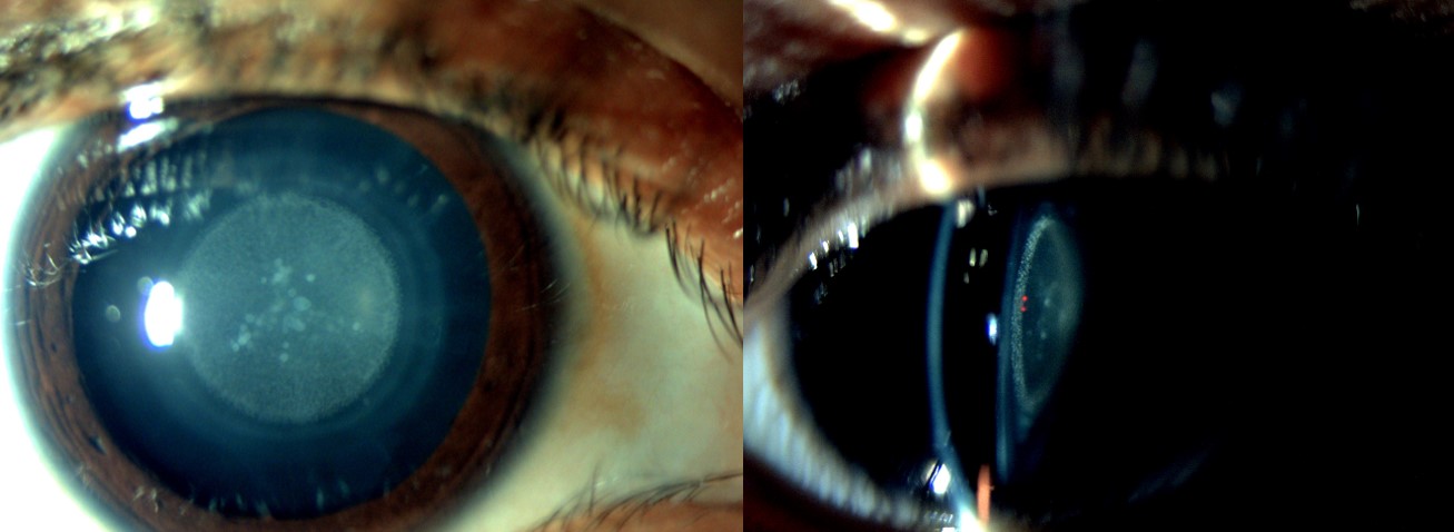
Slit-Lamp Examination of Pediatric Zonular Cataract. This zonular cataract exhibits diffuse and oblique illumination on slit-lamp examination
Contributed by Dr. Priyadarshi Gupta, MBBS, (MS) ophthalmology
(Click Image to Enlarge)
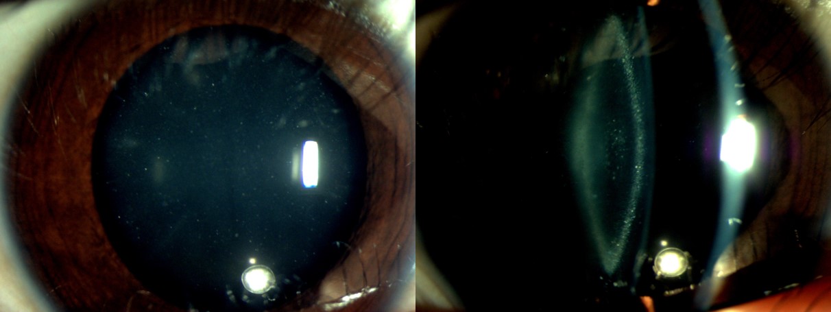
Pediatric Cataract, Zonular Type. This slit-lamp photograph depicts the zonular type of pediatric cataract with a blue dot cataract component.
Contributed by Dr. Priyadarshi Gupta, MBBS, (MS) Ophthalmology
(Click Image to Enlarge)
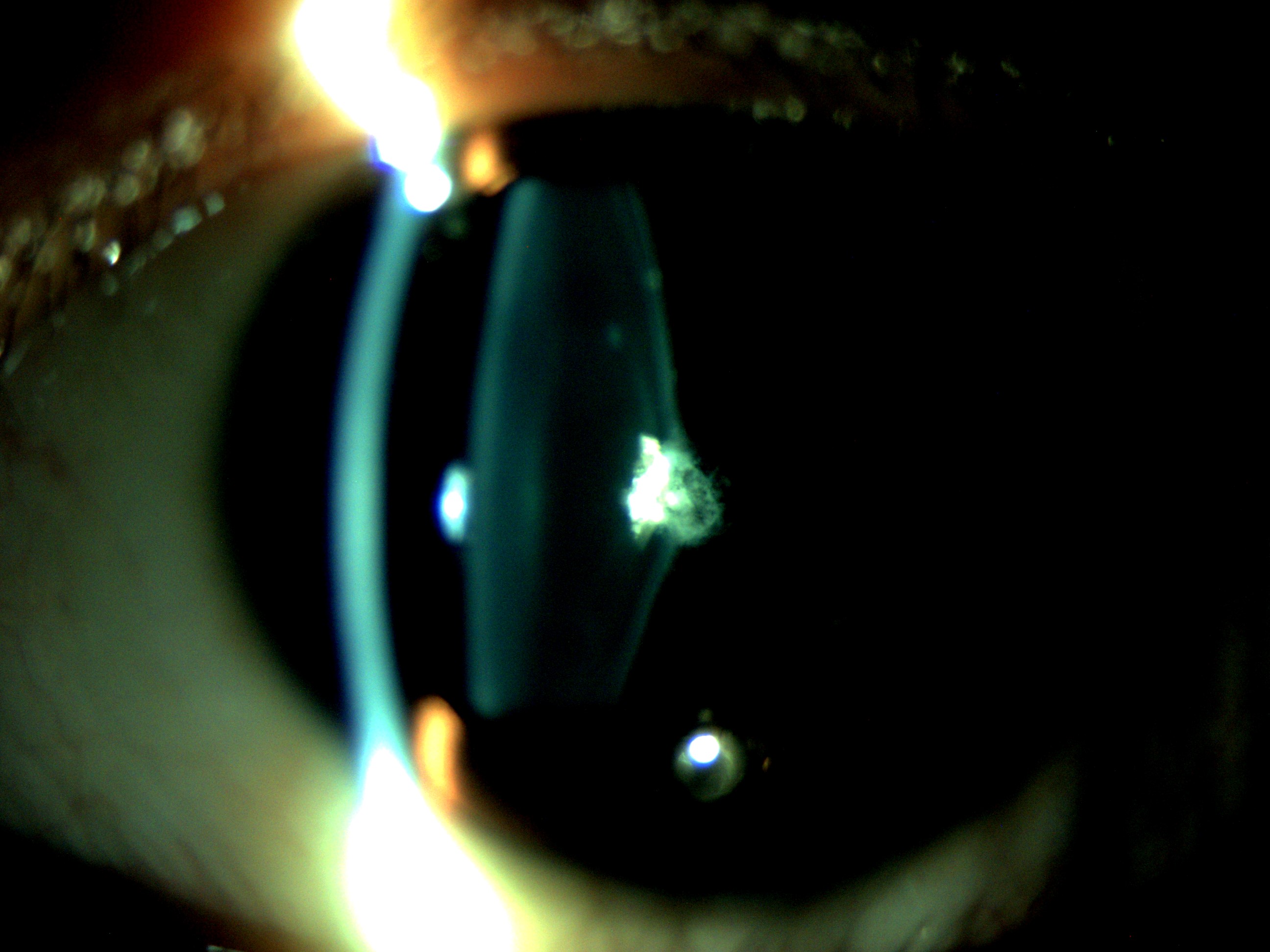
Pediatric Cataract, Posterior Lenticonus. Slit-lamp photograph showing a posterior lenticonus variant of pediatric cataract. Located near the eye's nodal point, posterior lenticonus typically impairs visual acuity more than anterior opacities. This form is frequently associated with Lowe syndrome.
Contributed by Dr. Priyadarshi Gupta, MBBS, (MS) Ophthalmology
(Click Image to Enlarge)
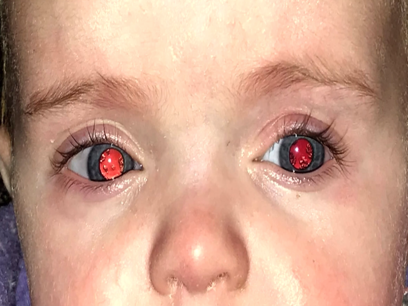
Congenital Cataracts and Abnormal Galactose Metabolism. Peripheral lenticular opacities of this type are often seen in patients with galactosemia or galactokinase deficiency.
Contributed by Prof. Bhupendra C. K. Patel MD, FRCS
(Click Image to Enlarge)

Red Reflex. This image shows a normal red reflex in each eye with equal clarity and brightness. The red reflex indicates the absence of opacification of the globe structures: cornea, aqueous, lens, vitreous, and a normal retina.
Contributed by Prof. Bhupendra C. K. Patel MD, FRCS
References
Khokhar SK, Pillay G, Dhull C, Agarwal E, Mahabir M, Aggarwal P. Pediatric cataract. Indian journal of ophthalmology. 2017 Dec:65(12):1340-1349. doi: 10.4103/ijo.IJO_1023_17. Epub [PubMed PMID: 29208814]
. Childhood cataract: magnitude, management, economics and impact. Community eye health. 2004:17(50):17-8 [PubMed PMID: 17491800]
Gilbert C, Foster A. Childhood blindness in the context of VISION 2020--the right to sight. Bulletin of the World Health Organization. 2001:79(3):227-32 [PubMed PMID: 11285667]
Solebo AL, Teoh L, Rahi J. Epidemiology of blindness in children. Archives of disease in childhood. 2017 Sep:102(9):853-857. doi: 10.1136/archdischild-2016-310532. Epub 2017 May 2 [PubMed PMID: 28465303]
Mendoza-Moreira AL, Voigt AM, Stingl JV, Rezapour J, Wagner FM, Schuster AK, Hoffmann EM. Paul Glaucoma Implant following Congenital Cataract Surgery in a Pediatric Cohort. Journal of clinical medicine. 2024 May 15:13(10):. doi: 10.3390/jcm13102914. Epub 2024 May 15 [PubMed PMID: 38792454]
Holmes JM, Leske DA, Burke JP, Hodge DO. Birth prevalence of visually significant infantile cataract in a defined U.S. population. Ophthalmic epidemiology. 2003 Apr:10(2):67-74 [PubMed PMID: 12660855]
Level 2 (mid-level) evidencePeake JN, Knowles RL, Shawe J, Rankin J, Copp AJ. Maternal ethnicity and the prevalence of British pregnancies affected by neural tube defects. Birth defects research. 2021 Jul 15:113(12):968-980. doi: 10.1002/bdr2.1893. Epub 2021 Mar 23 [PubMed PMID: 33754462]
Shiels A, Hejtmancik JF. Inherited cataracts: Genetic mechanisms and pathways new and old. Experimental eye research. 2021 Aug:209():108662. doi: 10.1016/j.exer.2021.108662. Epub 2021 Jun 12 [PubMed PMID: 34126080]
Shiels A, Hejtmancik JF. Mutations and mechanisms in congenital and age-related cataracts. Experimental eye research. 2017 Mar:156():95-102. doi: 10.1016/j.exer.2016.06.011. Epub 2016 Jun 19 [PubMed PMID: 27334249]
Self JE, Taylor R, Solebo AL, Biswas S, Parulekar M, Dev Borman A, Ashworth J, McClenaghan R, Abbott J, O'Flynn E, Hildebrand D, Lloyd IC. Cataract management in children: a review of the literature and current practice across five large UK centres. Eye (London, England). 2020 Dec:34(12):2197-2218. doi: 10.1038/s41433-020-1115-6. Epub 2020 Aug 10 [PubMed PMID: 32778738]
Al Shamrani M, Al Turkmani S. Update of intraocular lens implantation in children. Saudi journal of ophthalmology : official journal of the Saudi Ophthalmological Society. 2012 Jul:26(3):271-5. doi: 10.1016/j.sjopt.2012.05.005. Epub 2012 Jun 7 [PubMed PMID: 23961005]
Li Y, Tan Y, Xu C, Jin G, Chen H, Jin L, Luo L, Chen W, Lin H, Liu Y, Liu Z. Association Between Preoperative Ocular Parameters and Myopic Shift in Children Undergoing Primary Intraocular Lens Implantation. Translational vision science & technology. 2024 May 1:13(5):24. doi: 10.1167/tvst.13.5.24. Epub [PubMed PMID: 38809530]
Mets MB. Eye manifestations of intrauterine infections. Ophthalmology clinics of North America. 2001 Sep:14(3):521-31 [PubMed PMID: 11705152]
Ali A, Dockery PW, Downes DG, VanderVeen DK, Elhusseiny AM. Primary Suspect Drugs of Cataracts in Pediatric Patients: An FDA Adverse Events Reporting Database Analysis. Journal of cataract and refractive surgery. 2025 Jul 11:():. doi: 10.1097/j.jcrs.0000000000001738. Epub 2025 Jul 11 [PubMed PMID: 40643173]
Trumler AA. Evaluation of pediatric cataracts and systemic disorders. Current opinion in ophthalmology. 2011 Sep:22(5):365-79. doi: 10.1097/ICU.0b013e32834994dc. Epub [PubMed PMID: 21832913]
Level 3 (low-level) evidenceSantana A, Waiswo M. The genetic and molecular basis of congenital cataract. Arquivos brasileiros de oftalmologia. 2011 Mar-Apr:74(2):136-42 [PubMed PMID: 21779674]
Wadhwani M, Vashist P, Singh SS, Gupta V, Gupta N, Saxena R. Prevalence and causes of childhood blindness in India: A systematic review. Indian journal of ophthalmology. 2020 Feb:68(2):311-315. doi: 10.4103/ijo.IJO_2076_18. Epub [PubMed PMID: 31957718]
Level 1 (high-level) evidenceSheeladevi S, Lawrenson JG, Fielder AR, Suttle CM. Global prevalence of childhood cataract: a systematic review. Eye (London, England). 2016 Sep:30(9):1160-9. doi: 10.1038/eye.2016.156. Epub 2016 Aug 12 [PubMed PMID: 27518543]
Level 1 (high-level) evidenceTariq MA, Uddin QS, Ahmed B, Sheikh S, Ali U, Mohiuddin A. Prevalence of Pediatric Cataract in Asia: A Systematic Review and Meta-Analysis. Journal of current ophthalmology. 2022 Apr-Jun:34(2):148-159. doi: 10.4103/joco.joco_339_21. Epub 2022 Jul 26 [PubMed PMID: 36147271]
Level 1 (high-level) evidenceLin H, Yang Y, Chen J, Zhong X, Liu Z, Lin Z, Chen W, Luo L, Qu B, Zhang X, Zheng D, Zhan J, Wu H, Wang Z, Geng Y, Xiang W, Chen W, Liu Y, CCPMOH Study Group. Congenital cataract: prevalence and surgery age at Zhongshan Ophthalmic Center (ZOC). PloS one. 2014:9(7):e101781. doi: 10.1371/journal.pone.0101781. Epub 2014 Jul 3 [PubMed PMID: 24992190]
Carlson J, McBride K, O'Connor M. Drugs associated with cataract formation represent an unmet need in cataract research. Frontiers in medicine. 2022:9():947659. doi: 10.3389/fmed.2022.947659. Epub 2022 Aug 15 [PubMed PMID: 36045926]
Javadiyan S, Craig JE, Souzeau E, Sharma S, Lower KM, Mackey DA, Staffieri SE, Elder JE, Taranath D, Straga T, Black J, Pater J, Casey T, Hewitt AW, Burdon KP. High-Throughput Genetic Screening of 51 Pediatric Cataract Genes Identifies Causative Mutations in Inherited Pediatric Cataract in South Eastern Australia. G3 (Bethesda, Md.). 2017 Oct 5:7(10):3257-3268. doi: 10.1534/g3.117.300109. Epub 2017 Oct 5 [PubMed PMID: 28839118]
Level 3 (low-level) evidenceCvekl A, Duncan MK. Genetic and epigenetic mechanisms of gene regulation during lens development. Progress in retinal and eye research. 2007 Nov:26(6):555-97 [PubMed PMID: 17905638]
Level 3 (low-level) evidenceVerma IC, Paliwal P, Singh K. Genetic Testing in Pediatric Ophthalmology. Indian journal of pediatrics. 2018 Mar:85(3):228-236. doi: 10.1007/s12098-017-2453-7. Epub 2017 Oct 2 [PubMed PMID: 28971364]
Pollreisz A, Schmidt-Erfurth U. Diabetic cataract-pathogenesis, epidemiology and treatment. Journal of ophthalmology. 2010:2010():608751. doi: 10.1155/2010/608751. Epub 2010 Jun 17 [PubMed PMID: 20634936]
Lu B, Yang Y. Detection of TORCH pathogens in children with congenital cataracts. Experimental and therapeutic medicine. 2016 Aug:12(2):1159-1164 [PubMed PMID: 27446337]
Okoye GS, Gurnani B. Traumatic Cataract. StatPearls. 2025 Jan:(): [PubMed PMID: 37603642]
Böhm EW, Buonfiglio F, Voigt AM, Bachmann P, Safi T, Pfeiffer N, Gericke A. Oxidative stress in the eye and its role in the pathophysiology of ocular diseases. Redox biology. 2023 Dec:68():102967. doi: 10.1016/j.redox.2023.102967. Epub 2023 Nov 18 [PubMed PMID: 38006824]
Schmid PWN, Lim NCH, Peters C, Back KC, Bourgeois B, Pirolt F, Richter B, Peschek J, Puk O, Amarie OV, Dalke C, Haslbeck M, Weinkauf S, Madl T, Graw J, Buchner J. Imbalances in the eye lens proteome are linked to cataract formation. Nature structural & molecular biology. 2021 Feb:28(2):143-151. doi: 10.1038/s41594-020-00543-9. Epub 2021 Jan 11 [PubMed PMID: 33432246]
Theophanous CN, Wolfgeher DJ, Farooq AV, Hilkert Rodriguez S. Biomarkers of Pediatric Cataracts: A Proteomics Analysis of Aqueous Fluid. International journal of molecular sciences. 2023 May 20:24(10):. doi: 10.3390/ijms24109040. Epub 2023 May 20 [PubMed PMID: 37240389]
Shiels A, Hejtmancik JF. Biology of Inherited Cataracts and Opportunities for Treatment. Annual review of vision science. 2019 Sep 15:5():123-149. doi: 10.1146/annurev-vision-091517-034346. Epub [PubMed PMID: 31525139]
Stambolian D. Galactose and cataract. Survey of ophthalmology. 1988 Mar-Apr:32(5):333-49 [PubMed PMID: 3043741]
Level 3 (low-level) evidenceDezhagah H. Circular anterior lens capsule rupture caused by blunt ocular trauma. Middle East African journal of ophthalmology. 2010 Jan:17(1):103-5. doi: 10.4103/0974-9233.61227. Epub [PubMed PMID: 20543947]
James ER. The etiology of steroid cataract. Journal of ocular pharmacology and therapeutics : the official journal of the Association for Ocular Pharmacology and Therapeutics. 2007 Oct:23(5):403-20 [PubMed PMID: 17900234]
Gurcan MN, Boucheron LE, Can A, Madabhushi A, Rajpoot NM, Yener B. Histopathological image analysis: a review. IEEE reviews in biomedical engineering. 2009:2():147-71. doi: 10.1109/RBME.2009.2034865. Epub 2009 Oct 30 [PubMed PMID: 20671804]
Bornstein MH, Putnick DL, Suwalsky JT, Gini M. Maternal chronological age, prenatal and perinatal history, social support, and parenting of infants. Child development. 2006 Jul-Aug:77(4):875-92 [PubMed PMID: 16942495]
Bremond-Gignac D, Daruich A, Robert MP, Valleix S. Recent developments in the management of congenital cataract. Annals of translational medicine. 2020 Nov:8(22):1545. doi: 10.21037/atm-20-3033. Epub [PubMed PMID: 33313290]
Medsinge A, Nischal KK. Pediatric cataract: challenges and future directions. Clinical ophthalmology (Auckland, N.Z.). 2015:9():77-90. doi: 10.2147/OPTH.S59009. Epub 2015 Jan 7 [PubMed PMID: 25609909]
Level 3 (low-level) evidencePatil-Chhablani P, Kekunnaya R, Nischal KK. Complex Cases in Pediatric Cataract. Developments in ophthalmology. 2016:57():85-106. doi: 10.1159/000442505. Epub 2016 Apr 1 [PubMed PMID: 27043394]
Level 3 (low-level) evidenceBökenkamp A, Ludwig M. The oculocerebrorenal syndrome of Lowe: an update. Pediatric nephrology (Berlin, Germany). 2016 Dec:31(12):2201-2212 [PubMed PMID: 27011217]
Rajalakshmi AR, Rajeshwari M. Efficacy of Brückner's Test for Screening of Refractive Errors by Non-ophthalmologist Versus Ophthalmologist: A Comparative Study. Middle East African journal of ophthalmology. 2019 Oct-Dec:26(4):185-188. doi: 10.4103/meajo.MEAJO_121_19. Epub 2020 Jan 29 [PubMed PMID: 32153328]
Level 2 (mid-level) evidenceCotter SA, Tarczy-Hornoch K, Song E, Lin J, Borchert M, Azen SP, Varma R, Multi-Ethnic Pediatric Eye Disease Study Group. Fixation preference and visual acuity testing in a population-based cohort of preschool children with amblyopia risk factors. Ophthalmology. 2009 Jan:116(1):145-53. doi: 10.1016/j.ophtha.2008.08.031. Epub 2008 Oct 29 [PubMed PMID: 18962921]
Boop JJ, van Dalen JT, Tyner GS. Assessment of the Catford drum in visual acuity testing and its use as a measurement of visual performance in low-vision patients. The British journal of ophthalmology. 1987 Oct:71(10):797-802 [PubMed PMID: 3676152]
Caltrider D, Gupta A, Tripathy K. Evaluation of Visual Acuity. StatPearls. 2025 Jan:(): [PubMed PMID: 33231977]
Amaya L, Taylor D, Russell-Eggitt I, Nischal KK, Lengyel D. The morphology and natural history of childhood cataracts. Survey of ophthalmology. 2003 Mar-Apr:48(2):125-44 [PubMed PMID: 12686301]
Level 3 (low-level) evidenceZetterström C, Lundvall A, Kugelberg M. Cataracts in children. Journal of cataract and refractive surgery. 2005 Apr:31(4):824-40 [PubMed PMID: 15899463]
Braddick O, Atkinson J. Development of human visual function. Vision research. 2011 Jul 1:51(13):1588-609. doi: 10.1016/j.visres.2011.02.018. Epub 2011 Feb 26 [PubMed PMID: 21356229]
Papageorgiou E, McLean RJ, Gottlob I. Nystagmus in childhood. Pediatrics and neonatology. 2014 Oct:55(5):341-51. doi: 10.1016/j.pedneo.2014.02.007. Epub 2014 Jul 31 [PubMed PMID: 25086850]
Giangiacomo A, Beck A. Pediatric glaucoma: review of recent literature. Current opinion in ophthalmology. 2017 Mar:28(2):199-203. doi: 10.1097/ICU.0000000000000349. Epub [PubMed PMID: 27875350]
Level 3 (low-level) evidenceVasavada AR, Nihalani BR. Pediatric cataract surgery. Current opinion in ophthalmology. 2006 Feb:17(1):54-61 [PubMed PMID: 16436925]
Level 3 (low-level) evidenceVasavada V. Paradigms for Pediatric Cataract Surgery. Asia-Pacific journal of ophthalmology (Philadelphia, Pa.). 2018 Mar-Apr:7(2):123-127. doi: 10.22608/APO.2017202. Epub 2017 Sep 25 [PubMed PMID: 28971632]
Adams GGW. The Enduring Value of Newborn Red Reflex Testing as a Screening Tool. JAMA ophthalmology. 2021 Jan 1:139(1):40-41. doi: 10.1001/jamaophthalmol.2020.4853. Epub [PubMed PMID: 33180105]
Subhi Y, Schmidt DC, Al-Bakri M, Bach-Holm D, Kessel L. Diagnostic Test Accuracy of the Red Reflex Test for Ocular Pathology in Infants: A Meta-analysis. JAMA ophthalmology. 2021 Jan 1:139(1):33-40. doi: 10.1001/jamaophthalmol.2020.4854. Epub [PubMed PMID: 33180103]
Level 1 (high-level) evidenceSun M, Ma A, Li F, Cheng K, Zhang M, Yang H, Nie W, Zhao B. Sensitivity and Specificity of Red Reflex Test in Newborn Eye Screening. The Journal of pediatrics. 2016 Dec:179():192-196.e4. doi: 10.1016/j.jpeds.2016.08.048. Epub 2016 Sep 15 [PubMed PMID: 27640356]
Gurney SP, Makanjuola T, Kutubi M, Parulekar M, Abbott J. How to use…the direct ophthalmoscope. Archives of disease in childhood. Education and practice edition. 2018 Apr:103(2):102-109. doi: 10.1136/archdischild-2016-312378. Epub 2017 Sep 21 [PubMed PMID: 28935833]
American Academy of Pediatrics, Section on Ophthalmology, American Association for Pediatric Ophthalmology And Strabismus, American Academy of Ophthalmology, American Association of Certified Orthoptists. Red reflex examination in neonates, infants, and children. Pediatrics. 2008 Dec:122(6):1401-4. doi: 10.1542/peds.2008-2624. Epub [PubMed PMID: 19047263]
Toli A, Perente A, Labiris G. Evaluation of the red reflex: An overview for the pediatrician. World journal of methodology. 2021 Sep 20:11(5):263-277. doi: 10.5662/wjm.v11.i5.263. Epub 2021 Sep 20 [PubMed PMID: 34631483]
Level 3 (low-level) evidenceVillanelo F, Minogue PJ, Maripillán J, Reyna-Jeldes M, Jensen-Flores J, García IE, Beyer EC, Pérez-Acle T, Berthoud VM, Martínez AD. Connexin channels and hemichannels are modulated differently by charge reversal at residues forming the intracellular pocket. Biological research. 2024 May 23:57(1):31. doi: 10.1186/s40659-024-00501-5. Epub 2024 May 23 [PubMed PMID: 38783330]
Mannstadt M, Bilezikian JP, Thakker RV, Hannan FM, Clarke BL, Rejnmark L, Mitchell DM, Vokes TJ, Winer KK, Shoback DM. Hypoparathyroidism. Nature reviews. Disease primers. 2017 Aug 31:3():17055. doi: 10.1038/nrdp.2017.55. Epub 2017 Aug 31 [PubMed PMID: 28857066]
Drusin LM. Syphilis: clinical manifestations, diagnosis, and treatment. The Urologic clinics of North America. 1984 Feb:11(1):121-30 [PubMed PMID: 6369700]
Jyoti M, Shirke S, Matalia H. Congenital rubella syndrome: Global issue. Journal of cataract and refractive surgery. 2015 May:41(5):1127. doi: 10.1016/j.jcrs.2015.04.021. Epub [PubMed PMID: 26049858]
Anderson S. GALT Deficiency Galactosemia. MCN. The American journal of maternal child nursing. 2018 Jan/Feb:43(1):44-51. doi: 10.1097/NMC.0000000000000388. Epub [PubMed PMID: 29215423]
Savige J, Sheth S, Leys A, Nicholson A, Mack HG, Colville D. Ocular features in Alport syndrome: pathogenesis and clinical significance. Clinical journal of the American Society of Nephrology : CJASN. 2015 Apr 7:10(4):703-9. doi: 10.2215/CJN.10581014. Epub 2015 Feb 3 [PubMed PMID: 25649157]
Level 3 (low-level) evidenceCarrillo-Carrasco N, Chandler RJ, Venditti CP. Combined methylmalonic acidemia and homocystinuria, cblC type. I. Clinical presentations, diagnosis and management. Journal of inherited metabolic disease. 2012 Jan:35(1):91-102. doi: 10.1007/s10545-011-9364-y. Epub 2011 Jul 12 [PubMed PMID: 21748409]
Ferenci P. Diagnosis of Wilson disease. Handbook of clinical neurology. 2017:142():171-180. doi: 10.1016/B978-0-444-63625-6.00014-8. Epub [PubMed PMID: 28433100]
Kaya A. Preoperative usage of ultrasound biomicroscopy in pediatric cataract. Arquivos brasileiros de oftalmologia. 2016 Feb:79(1):62. doi: 10.5935/0004-2749.20160019. Epub [PubMed PMID: 26840173]
Khokhar S, Tejwani LK, Kumar G, Kushmesh R. Approach to cataract with persistent hyperplastic primary vitreous. Journal of cataract and refractive surgery. 2011 Aug:37(8):1382-5. doi: 10.1016/j.jcrs.2011.06.012. Epub [PubMed PMID: 21782083]
Level 3 (low-level) evidenceCapozzi P, Morini C, Piga S, Cuttini M, Vadalà P. Corneal curvature and axial length values in children with congenital/infantile cataract in the first 42 months of life. Investigative ophthalmology & visual science. 2008 Nov:49(11):4774-8. doi: 10.1167/iovs.07-1564. Epub 2008 May 23 [PubMed PMID: 18502997]
Level 2 (mid-level) evidenceMoshirfar M, Buckner B, Ronquillo YC, Hofstedt D. Biometry in cataract surgery: a review of the current literature. Current opinion in ophthalmology. 2019 Jan:30(1):9-12. doi: 10.1097/ICU.0000000000000536. Epub [PubMed PMID: 30394990]
Level 3 (low-level) evidenceTrivedi RH, Wilson ME. Keratometry in pediatric eyes with cataract. Archives of ophthalmology (Chicago, Ill. : 1960). 2008 Jan:126(1):38-42. doi: 10.1001/archophthalmol.2007.22. Epub [PubMed PMID: 18195216]
Level 2 (mid-level) evidenceDavison JE. Eye involvement in inherited metabolic disorders. Therapeutic advances in ophthalmology. 2020 Jan-Dec:12():2515841420979109. doi: 10.1177/2515841420979109. Epub 2020 Dec 29 [PubMed PMID: 33447730]
Level 3 (low-level) evidenceBoniquet-Sanchez S, Sabater-Cruz N. Current Management of Amblyopia with New Technologies for Binocular Treatment. Vision (Basel, Switzerland). 2021 Jun 10:5(2):. doi: 10.3390/vision5020031. Epub 2021 Jun 10 [PubMed PMID: 34200969]
Heus P, Verbeek JH, Tikka C. Optical correction of refractive error for preventing and treating eye symptoms in computer users. The Cochrane database of systematic reviews. 2018 Apr 10:4(4):CD009877. doi: 10.1002/14651858.CD009877.pub2. Epub 2018 Apr 10 [PubMed PMID: 29633784]
Level 1 (high-level) evidenceAger L. Optical services for visually impaired children. Community eye health. 1998:11(27):38-40 [PubMed PMID: 17492038]
K Lodhi SA, Ramsali MV, Kulkarni DK, Surender P, Murty S. Safety of tropicamide and phenylephrine in pupillary mydriasis for cataract surgery. Saudi journal of ophthalmology : official journal of the Saudi Ophthalmological Society. 2021 Apr-Jun:35(2):108-111. doi: 10.4103/1319-4534.337859. Epub 2022 Feb 18 [PubMed PMID: 35391812]
Hamm LM, Boluk KA, Black JM, Dai S, Thompson B. Phenomenological approach to childhood cataract treatment in New Zealand using semi-structured interviews: how might we improve provision of care. BMJ open. 2019 Jan 28:9(1):e024869. doi: 10.1136/bmjopen-2018-024869. Epub 2019 Jan 28 [PubMed PMID: 30782745]
Level 3 (low-level) evidenceGlynn RJ, Rosner B, Christen WG. Evaluation of risk factors for cataract types in a competing risks framework. Ophthalmic epidemiology. 2009 Mar-Apr:16(2):98-106. doi: 10.1080/09286580902737532. Epub [PubMed PMID: 19353398]
Repka MX, Sutherland DR, Hatt SR, Rahmani B, Leske DA, Bothun ED, Young MP, Strominger MB, Tamkins SM, Beaulieu WT, Kraker RT, Holmes JM, Cotter SA, Pediatric Eye Disease Investigator Group. Effects of Age at Surgery and Laterality of Cataract on Visual Acuity 5 years After Surgery in Infants Left Aphakic. Ophthalmology. 2025 Jun 27:():. pii: S0161-6420(25)00393-8. doi: 10.1016/j.ophtha.2025.06.022. Epub 2025 Jun 27 [PubMed PMID: 40582417]
Infant Aphakia Treatment Study Group, Lambert SR, Buckley EG, Drews-Botsch C, DuBois L, Hartmann E, Lynn MJ, Plager DA, Wilson ME. The infant aphakia treatment study: design and clinical measures at enrollment. Archives of ophthalmology (Chicago, Ill. : 1960). 2010 Jan:128(1):21-7. doi: 10.1001/archophthalmol.2009.350. Epub [PubMed PMID: 20065212]
Level 1 (high-level) evidenceMagnusson G, Gyllén J, Haargaard B, Nyström A, Rosensvärd A, Scurei C, Kjellström U, Tornqvist K. The prevalence of visual axis opacification in the Swedish Pediatric Cataract Register. Acta ophthalmologica. 2024 Sep:102(6):e873-e882. doi: 10.1111/aos.16630. Epub 2024 Jan 22 [PubMed PMID: 38251769]
Lim ME, Buckley EG, Prakalapakorn SG. Update on congenital cataract surgery management. Current opinion in ophthalmology. 2017 Jan:28(1):87-92 [PubMed PMID: 27653605]
Level 3 (low-level) evidenceLin AA, Buckley EG. Update on pediatric cataract surgery and intraocular lens implantation. Current opinion in ophthalmology. 2010 Jan:21(1):55-9. doi: 10.1097/ICU.0b013e32833383cb. Epub [PubMed PMID: 19855277]
Level 3 (low-level) evidenceDevebacak A, Biler ED, Degirmenci C, Uretmen O. Optic Capture Without Anterior Vitrectomy in Pediatric Cataract Surgery. American journal of ophthalmology. 2023 Mar:247():88-95. doi: 10.1016/j.ajo.2022.11.007. Epub 2022 Nov 12 [PubMed PMID: 36375587]
Kohnen T, Davidova P, Lambert M, Wenner Y, Zubcov AA. Posterior continuous curvilinear capsulorhexis with anterior vitrectomy vs optic capture buttonholing without anterior vitrectomy in pediatric cataract surgery. Journal of cataract and refractive surgery. 2022 Jul 1:48(7):831-837. doi: 10.1097/j.jcrs.0000000000000846. Epub [PubMed PMID: 34670945]
Sukhija J, Kaur S, Korla S, Kumari K. Surgical challenges of posterior optic capture in pediatric cataract surgery. Indian journal of ophthalmology. 2024 Jan 1:72(1):51-55. doi: 10.4103/IJO.IJO_506_23. Epub 2023 Dec 22 [PubMed PMID: 38131569]
Eibschitz-Tsimhoni M, Archer SM, Del Monte MA. Intraocular lens power calculation in children. Survey of ophthalmology. 2007 Sep-Oct:52(5):474-82 [PubMed PMID: 17719370]
Level 3 (low-level) evidenceVasavada V, Shah SK, Vasavada VA, Vasavada AR, Trivedi RH, Srivastava S, Vasavada SA. Comparison of IOL power calculation formulae for pediatric eyes. Eye (London, England). 2016 Sep:30(9):1242-50. doi: 10.1038/eye.2016.171. Epub 2016 Aug 5 [PubMed PMID: 27494083]
Dahan E, Drusedau MU. Choice of lens and dioptric power in pediatric pseudophakia. Journal of cataract and refractive surgery. 1997:23 Suppl 1():618-23 [PubMed PMID: 9278814]
Level 2 (mid-level) evidenceEnyedi LB, Peterseim MW, Freedman SF, Buckley EG. Refractive changes after pediatric intraocular lens implantation. American journal of ophthalmology. 1998 Dec:126(6):772-81 [PubMed PMID: 9860000]
Oshika T, Nishina S, Ura S, Miyagi M, Nomura K, Mori T, Matsuki N, Endo T, Kurosaka D, Negishi K, Yoshida S, Nagamoto T. Long-Term Refractive Changes After Pediatric Cataract Surgery: An Average Follow-up of 15 Years. American journal of ophthalmology. 2025 May 28:277():305-312. doi: 10.1016/j.ajo.2025.05.035. Epub 2025 May 28 [PubMed PMID: 40447247]
Lenhart PD, Lambert SR. Current management of infantile cataracts. Survey of ophthalmology. 2022 Sep-Oct:67(5):1476-1505. doi: 10.1016/j.survophthal.2022.03.005. Epub 2022 Mar 17 [PubMed PMID: 35307324]
Level 3 (low-level) evidenceZhang JH, Ramke J, Lee CN, Gordon I, Safi S, Lingham G, Evans JR, Keel S. A Systematic Review of Clinical Practice Guidelines for Cataract: Evidence to Support the Development of the WHO Package of Eye Care Interventions. Vision (Basel, Switzerland). 2022 Jun 20:6(2):. doi: 10.3390/vision6020036. Epub 2022 Jun 20 [PubMed PMID: 35737423]
Level 1 (high-level) evidenceBell SJ, Oluonye N, Harding P, Moosajee M. Congenital cataract: a guide to genetic and clinical management. Therapeutic advances in rare disease. 2020 Jan-Dec:1():2633004020938061. doi: 10.1177/2633004020938061. Epub 2020 Jul 22 [PubMed PMID: 37180497]
Level 3 (low-level) evidenceGera T, Shah D, Garner P, Richardson M, Sachdev HS. Integrated management of childhood illness (IMCI) strategy for children under five. The Cochrane database of systematic reviews. 2016 Jun 22:2016(6):CD010123. doi: 10.1002/14651858.CD010123.pub2. Epub 2016 Jun 22 [PubMed PMID: 27378094]
Level 1 (high-level) evidenceAntwi-Adjei EK, Owusu E, Kobia-Acquah E, Dadzie EE, Anarfi E, Wanye S. Evaluation of postoperative refractive error correction after cataract surgery. PloS one. 2021:16(6):e0252787. doi: 10.1371/journal.pone.0252787. Epub 2021 Jun 17 [PubMed PMID: 34138900]
Ghasemi Fard F, Mirzaie H, Hosseini SA, Riazi A, Ebadi A. Vision-related tasks in children with visual impairment: a multi-method study. Frontiers in psychology. 2023:14():1180669. doi: 10.3389/fpsyg.2023.1180669. Epub 2023 Jul 13 [PubMed PMID: 37519380]
Chen AM, Cotter SA. The Amblyopia Treatment Studies: Implications for Clinical Practice. Advances in ophthalmology and optometry. 2016 Aug:1(1):287-305. doi: 10.1016/j.yaoo.2016.03.007. Epub [PubMed PMID: 28435934]
Level 3 (low-level) evidenceIsrar M, Zahir KK, Khattak A, Khattak IU, Gul N. Etiology of white pupillary reflex in pediatric age group. Romanian journal of ophthalmology. 2022 Jan-Mar:66(1):32-35. doi: 10.22336/rjo.2022.8. Epub [PubMed PMID: 35531443]
Prakhunhungsit S, Berrocal AM. Diagnostic and Management Strategies in Patients with Persistent Fetal Vasculature: Current Insights. Clinical ophthalmology (Auckland, N.Z.). 2020:14():4325-4335. doi: 10.2147/OPTH.S236117. Epub 2020 Dec 10 [PubMed PMID: 33335385]
Cheng KP, Hiles DA, Biglan AW. The differential diagnosis of leukokoria. Pediatric annals. 1990 Jun:19(6):376-83, 386 [PubMed PMID: 2200999]
Kembhavi SA, Sable N, Vora T, Arora B. Leukokoria: All That's White Is Not Retinoblastoma. Journal of clinical oncology : official journal of the American Society of Clinical Oncology. 2011 Jul 1:29(19):e586-7. doi: 10.1200/JCO.2011.34.8334. Epub 2011 Apr 18 [PubMed PMID: 21502547]
Level 3 (low-level) evidenceGurnani B, Kaur K. Re: Dot et al.: Incidence of retinal detachment, macular edema, and ocular hypertension after neodymium:yttrium-aluminum-garnet capsulotomy: a population-based nationwide study-The French YAG 2 Study (Ophthalmology. 2022;130:478-487). Ophthalmology. 2023 Dec:130(12):e43. doi: 10.1016/j.ophtha.2023.08.012. Epub 2023 Sep 20 [PubMed PMID: 37737811]
Kaur K, Zeppieri M, Gurnani B. Primary Congenital Glaucoma. StatPearls. 2025 Jan:(): [PubMed PMID: 34662067]
Paulbuddhe V, Addya S, Gurnani B, Singh D, Tripathy K, Chawla R. Sympathetic Ophthalmia: Where Do We Currently Stand on Treatment Strategies? Clinical ophthalmology (Auckland, N.Z.). 2021:15():4201-4218. doi: 10.2147/OPTH.S289688. Epub 2021 Oct 20 [PubMed PMID: 34707340]
Kaur K, Gurnani B. Microspherophakia. StatPearls. 2025 Jan:(): [PubMed PMID: 35015420]
Vohra V, Tuteja S, Gurnani B, Chawla H. Collagen Cross Linking for Keratoconus. StatPearls. 2025 Jan:(): [PubMed PMID: 32965942]
Kaur K, Gurnani B. Lenticonus. StatPearls. 2025 Jan:(): [PubMed PMID: 36943989]
Gurnani B, Kaur K, Sekaran S. First case of coloboma, lens neovascularization, traumatic cataract, and retinal detachment in a young Asian female. Clinical case reports. 2021 Sep:9(9):e04743. doi: 10.1002/ccr3.4743. Epub 2021 Aug 30 [PubMed PMID: 34484773]
Level 3 (low-level) evidenceAhmad B, Gurnani B, Patel BC. Herpes Simplex Keratitis. StatPearls. 2025 Jan:(): [PubMed PMID: 31424862]
Michael R, Bron AJ. The ageing lens and cataract: a model of normal and pathological ageing. Philosophical transactions of the Royal Society of London. Series B, Biological sciences. 2011 Apr 27:366(1568):1278-92. doi: 10.1098/rstb.2010.0300. Epub [PubMed PMID: 21402586]
Shandiz JH, Derakhshan A, Daneshyar A, Azimi A, Moghaddam HO, Yekta AA, Yazdi SH, Esmaily H. Effect of cataract type and severity on visual acuity and contrast sensitivity. Journal of ophthalmic & vision research. 2011 Jan:6(1):26-31 [PubMed PMID: 22454703]
Mencucci R, Stefanini S, Favuzza E, Cennamo M, De Vitto C, Mossello E. Beyond vision:Cataract and health status in old age, a narrative review. Frontiers in medicine. 2023:10():1110383. doi: 10.3389/fmed.2023.1110383. Epub 2023 Mar 16 [PubMed PMID: 37007780]
Level 3 (low-level) evidenceVijayalakshmi P, Njambi L. Paediatric cataract: challenges and complications. Community eye health. 2016:29(94):34-35 [PubMed PMID: 27833263]
Kalia A, Gandhi T, Chatterjee G, Swami P, Dhillon H, Bi S, Chauhan N, Gupta SD, Sharma P, Sood S, Ganesh S, Mathur U, Sinha P. Assessing the impact of a program for late surgical intervention in early-blind children. Public health. 2017 May:146():15-23. doi: 10.1016/j.puhe.2016.12.036. Epub 2017 Feb 1 [PubMed PMID: 28404468]
Sen S, Singh P, Saxena R. Management of amblyopia in pediatric patients: Current insights. Eye (London, England). 2022 Jan:36(1):44-56. doi: 10.1038/s41433-021-01669-w. Epub 2021 Jul 7 [PubMed PMID: 34234293]
Long V, Chen S, Hatt S. Surgical interventions for bilateral congenital cataract. The Cochrane database of systematic reviews. 2006 Jul 19:2006(3):CD003171 [PubMed PMID: 16855998]
Level 1 (high-level) evidenceWagner IV, Stewart MW, Dorairaj SK. Updates on the Diagnosis and Management of Glaucoma. Mayo Clinic proceedings. Innovations, quality & outcomes. 2022 Dec:6(6):618-635. doi: 10.1016/j.mayocpiqo.2022.09.007. Epub 2022 Nov 16 [PubMed PMID: 36405987]
Level 2 (mid-level) evidenceShrestha UD, Shrestha MK. Visual Axis Opacification in Children Following Paediatric Cataract Surgery. JNMA; journal of the Nepal Medical Association. 2014 Oct-Dec:52(196):1024-30 [PubMed PMID: 26982905]
Khaja WA, Verma M, Shoss BL, Yen KG. Visual axis opacification in children. Ophthalmology. 2011 Jan:118(1):224-5. doi: 10.1016/j.ophtha.2010.08.030. Epub [PubMed PMID: 21199720]
Level 3 (low-level) evidenceBaden C, Shija F, Lewallen S, Courtright P, Hall A. Glaucoma after pediatric cataract surgery in a population with limited access to care. Journal of AAPOS : the official publication of the American Association for Pediatric Ophthalmology and Strabismus. 2013 Apr:17(2):158-62. doi: 10.1016/j.jaapos.2012.11.009. Epub 2013 Mar 22 [PubMed PMID: 23528376]
Level 2 (mid-level) evidenceWhitman MC, Vanderveen DK. Complications of pediatric cataract surgery. Seminars in ophthalmology. 2014 Sep-Nov:29(5-6):414-20. doi: 10.3109/08820538.2014.959192. Epub [PubMed PMID: 25325868]
Cruz OA, Repka MX, Hercinovic A, Cotter SA, Lambert SR, Hutchinson AK, Sprunger DT, Morse CL, Wallace DK, American Academy of Ophthalmology Preferred Practice Pattern Pediatric Ophthalmology/Strabismus Panel. Amblyopia Preferred Practice Pattern. Ophthalmology. 2023 Mar:130(3):P136-P178. doi: 10.1016/j.ophtha.2022.11.003. Epub 2022 Dec 14 [PubMed PMID: 36526450]
Konopińska J, Młynarczyk M, Dmuchowska DA, Obuchowska I. Posterior Capsule Opacification: A Review of Experimental Studies. Journal of clinical medicine. 2021 Jun 27:10(13):. doi: 10.3390/jcm10132847. Epub 2021 Jun 27 [PubMed PMID: 34199147]
Kanukollu VM, Sood G. Strabismus. StatPearls. 2025 Jan:(): [PubMed PMID: 32809617]
Althiabi S, Aljbreen AJ, Alshutily A, Althwiny FA. Postoperative Endophthalmitis After Cataract Surgery: An Update. Cureus. 2022 Feb:14(2):e22003. doi: 10.7759/cureus.22003. Epub 2022 Feb 8 [PubMed PMID: 35340495]
Jalali S. Retinal detachment. Community eye health. 2003:16(46):25-6 [PubMed PMID: 17491854]
Schiefer U, Kraus C, Baumbach P, Ungewiß J, Michels R. Refractive errors. Deutsches Arzteblatt international. 2016 Oct 14:113(41):693-702. doi: 10.3238/arztebl.2016.0693. Epub [PubMed PMID: 27839543]
Tripathy K, Chawla R, Temkar S, Sagar P, Kashyap S, Pushker N, Sharma YR. Phthisis Bulbi-a Clinicopathological Perspective. Seminars in ophthalmology. 2018:33(6):788-803. doi: 10.1080/08820538.2018.1477966. Epub 2018 Jun 14 [PubMed PMID: 29902388]
Level 3 (low-level) evidenceGasper C, Trivedi RH, Wilson ME. Complications of Pediatric Cataract Surgery. Developments in ophthalmology. 2016:57():69-84. doi: 10.1159/000442502. Epub 2016 Apr 1 [PubMed PMID: 27043393]
Repka MX. Visual Rehabilitation in Pediatric Aphakia. Developments in ophthalmology. 2016:57():49-68. doi: 10.1159/000442501. Epub 2016 Apr 1 [PubMed PMID: 27043392]
Vagge A, Nelson LB. Amblyopia update: new treatments. Current opinion in ophthalmology. 2016 Sep:27(5):380-6. doi: 10.1097/ICU.0000000000000293. Epub [PubMed PMID: 27152486]
Level 3 (low-level) evidenceKaur K, Gurnani B. Low Vision Aids. StatPearls. 2025 Jan:(): [PubMed PMID: 36256771]
Al-Essa RS, Alfawaz AM. New insights into cataract surgery in patients with uveitis: A detailed review of the current literature. Saudi journal of ophthalmology : official journal of the Saudi Ophthalmological Society. 2022 Apr-Jun:36(2):133-141. doi: 10.4103/sjopt.sjopt_147_21. Epub 2022 Feb 18 [PubMed PMID: 36211311]
Hutchinson AK, Morse CL, Hercinovic A, Cruz OA, Sprunger DT, Repka MX, Lambert SR, Wallace DK, American Academy of Ophthalmology Preferred Practice Pattern Pediatric Ophthalmology/Strabismus Panel. Pediatric Eye Evaluations Preferred Practice Pattern. Ophthalmology. 2023 Mar:130(3):P222-P270. doi: 10.1016/j.ophtha.2022.10.030. Epub 2022 Dec 19 [PubMed PMID: 36543602]
de Zárate BR, Tejedor J. Current concepts in the management of amblyopia. Clinical ophthalmology (Auckland, N.Z.). 2007 Dec:1(4):403-14 [PubMed PMID: 19668517]
Tsai LT, Hsu JL, Wu CT, Chen CC, Su YC. A New Visual Stimulation Program for Improving Visual Acuity in Children with Visual Impairment: A Pilot Study. Frontiers in human neuroscience. 2016:10():157. doi: 10.3389/fnhum.2016.00157. Epub 2016 Apr 18 [PubMed PMID: 27148014]
Level 3 (low-level) evidenceDagg W, Forgeron P, Macartney G, Chartrand J. Parents' management of adolescent patients' postoperative pain after discharge: A qualitative study. Canadian journal of pain = Revue canadienne de la douleur. 2020 Sep 24:4(3):51-60. doi: 10.1080/24740527.2020.1783524. Epub 2020 Sep 24 [PubMed PMID: 33987511]
Level 2 (mid-level) evidenceAdeniyi YC, Omigbodun OO. Effect of a classroom-based intervention on the social skills of pupils with intellectual disability in Southwest Nigeria. Child and adolescent psychiatry and mental health. 2016:10(1):29. doi: 10.1186/s13034-016-0118-3. Epub 2016 Sep 2 [PubMed PMID: 27594901]
Alizary A, Ahmad K, Al Bakri A. Parental experience with an ocular genetic counseling services in Saudi Arabia. Saudi journal of ophthalmology : official journal of the Saudi Ophthalmological Society. 2023 Oct-Dec:37(4):296-300. doi: 10.4103/sjopt.sjopt_154_23. Epub 2023 Oct 24 [PubMed PMID: 38155671]
Lewis H, James I. Update on anaesthesia for paediatric ophthalmic surgery. BJA education. 2021 Jan:21(1):32-38. doi: 10.1016/j.bjae.2020.09.002. Epub 2020 Nov 5 [PubMed PMID: 33456972]
Vijayalakshmi P, Njambi L. Postoperative care for paediatric cataract patients. Community eye health. 2016:29(94):32-34 [PubMed PMID: 27833262]
Rezaie L, Kendi S. Exploration of the Influential Factors on Adherence to Occupational Therapy in Parents of Children with Cerebral Palsy: A Qualitative Study. Patient preference and adherence. 2020:14():63-72. doi: 10.2147/PPA.S229535. Epub 2020 Jan 13 [PubMed PMID: 32021118]
Level 2 (mid-level) evidenceSingh T, Otero CE, Li K, Valencia SM, Nelson AN, Permar SR. Vaccines for Perinatal and Congenital Infections-How Close Are We? Frontiers in pediatrics. 2020:8():569. doi: 10.3389/fped.2020.00569. Epub 2020 Dec 15 [PubMed PMID: 33384972]
Malik AN, Evans JR, Gupta S, Mariotti S, Gordon I, Bowman R, Gilbert C. Universal newborn eye screening: a systematic review of the literature and review of international guidelines. Journal of global health. 2022 Oct 21:12():12003. doi: 10.7189/jogh.12.12003. Epub 2022 Oct 21 [PubMed PMID: 36269293]
Level 1 (high-level) evidenceBadiu Tișa I, Achim AC, Cozma-Petruț A. The Importance of Neonatal Screening for Galactosemia. Nutrients. 2022 Dec 20:15(1):. doi: 10.3390/nu15010010. Epub 2022 Dec 20 [PubMed PMID: 36615667]
MacLaren Chorney J, Twycross A, Mifflin K, Archibald K. Can we improve parents' management of their children's postoperative pain at home? Pain research & management. 2014 Jul-Aug:19(4):e115-23 [PubMed PMID: 25106030]
Gyllén J, Magnusson G, Forsberg A. Parents' Reported Experiences When Having a Child with Cataract-Important Aspects of Self-Management Obtained from the Paediatric Cataract Register (PECARE). International journal of environmental research and public health. 2020 Aug 31:17(17):. doi: 10.3390/ijerph17176329. Epub 2020 Aug 31 [PubMed PMID: 32878121]
Bhattad PB, Pacifico L. Empowering Patients: Promoting Patient Education and Health Literacy. Cureus. 2022 Jul:14(7):e27336. doi: 10.7759/cureus.27336. Epub 2022 Jul 27 [PubMed PMID: 36043002]
Macias AA, Goldfarb J. Preoperative Evaluation of Patients Undergoing Eye Surgery. Anesthesiology clinics. 2024 Mar:42(1):159-168. doi: 10.1016/j.anclin.2023.08.006. Epub 2023 Sep 25 [PubMed PMID: 38278587]
Sen P, Gupta N, Mohan A, Shah C, Sen A, Jain E. Causes of delayed presentation of pediatric cataract: A questionnaire-based prospective study at a tertiary eye care center in central rural India. Indian journal of ophthalmology. 2020 Apr:68(4):603-607. doi: 10.4103/ijo.IJO_872_19. Epub [PubMed PMID: 32174578]
Moshirfar M, Churgin DS, Hsu M. Femtosecond laser-assisted cataract surgery: a current review. Middle East African journal of ophthalmology. 2011 Oct:18(4):285-91. doi: 10.4103/0974-9233.90129. Epub [PubMed PMID: 22224016]
Katre D, Selukar K. The Prevalence of Cataract in Children. Cureus. 2022 Oct:14(10):e30135. doi: 10.7759/cureus.30135. Epub 2022 Oct 10 [PubMed PMID: 36381901]
Hall DE, Prochazka AV, Fink AS. Informed consent for clinical treatment. CMAJ : Canadian Medical Association journal = journal de l'Association medicale canadienne. 2012 Mar 20:184(5):533-40. doi: 10.1503/cmaj.112120. Epub 2012 Mar 5 [PubMed PMID: 22392947]
Martínez-Fernández MC, Castiñeiras-Martín S, Liébana-Presa C, Fernández-Martínez E, Gomes L, Marques-Sanchez P. SBAR Method for Improving Well-Being in the Internal Medicine Unit: Quasi-Experimental Research. International journal of environmental research and public health. 2022 Dec 14:19(24):. doi: 10.3390/ijerph192416813. Epub 2022 Dec 14 [PubMed PMID: 36554696]
Moeenuddin Z, Kim-Kupfer C, Owchar E, Baker J, Duffield A, Santoro T. The Influence of Care Coordination on Patients With Special Health Care Needs in a Pediatric Residency Continuity Clinic. Global pediatric health... 2019:6():2333794X19848677. doi: 10.1177/2333794X19848677. Epub 2019 May 8 [PubMed PMID: 31106252]
Edgman-Levitan S, Schoenbaum SC. Patient-centered care: achieving higher quality by designing care through the patient's eyes. Israel journal of health policy research. 2021 Mar 5:10(1):21. doi: 10.1186/s13584-021-00459-9. Epub 2021 Mar 5 [PubMed PMID: 33673875]
Level 2 (mid-level) evidenceForster AJ, Dervin G, Martin C, Papp S. Improving patient safety through the systematic evaluation of patient outcomes. Canadian journal of surgery. Journal canadien de chirurgie. 2012 Dec:55(6):418-25. doi: 10.1503/cjs.007811. Epub [PubMed PMID: 23177520]
Level 1 (high-level) evidenceWu S, Yan X, Wu W, Hou L, Liang Y, Luo X. Early juvenile cataract in newly diagnosed type 1 diabetic patients: a description of two cases. Journal of pediatric endocrinology & metabolism : JPEM. 2024 Jul 26:37(7):652-656. doi: 10.1515/jpem-2024-0005. Epub 2024 May 31 [PubMed PMID: 38814569]
Level 3 (low-level) evidence