 Anatomy, Bony Pelvis and Lower Limb: Arches of the Foot
Introduction
An arched foot is a distinctive feature of humans, setting them apart from other primates. The foot arches contribute to upright posture and facilitate weight bearing. The human foot contains 2 longitudinal arches, medial and lateral, and a transverse arch with anterior and posterior components that function cohesively (see Image. Bones Forming the Foot Arches).[1][2] The tarsals and metatarsals form the arches of the foot. Ligaments, along with extrinsic and intrinsic muscles, provide additional support and stability. Of the 2 longitudinal arches, the medial arch is higher. Of the transverse arch segments, the anterior portion is complete, whereas the posterior region is half-dome-shaped and incomplete. These arches render the sole concave, producing an incomplete footprint. Arches are present from birth but may be obscured in children by the subcutaneous fat pad.
The foot arches provide a flexible platform for weight support and act as a multisegmented lever during walking, running, and jumping. An arched foot absorbs shock and adapts to uneven surfaces. Deformities may result from congenital anomalies or acquired conditions, including improper footwear or prolonged use of high heels.[3][4] Understanding foot arch anatomy and function helps prevent and manage injuries related to abnormal weight distribution and gait patterns.
Structure and Function
Register For Free And Read The Full Article
 Search engine and full access to all medical articles
- 10 free questions in your specialty
- Free CME/CE Activities
- Free daily question in your email
- Save favorite articles to your dashboard
- Emails offering discounts
Learn more about a Subscription to StatPearls Point-of-Care
Structure and Function
Classification of the Foot Arches
The human foot features 2 longitudinal arches, medial and lateral, and a transverse arch comprising anterior and posterior segments. The arches are often compared to a stone bridge with 2 ends, pillars, a summit, and a keystone, supported by mechanisms analogous to intersegmental ties (staples), tie-beams, and slings (see Image. Stone Bridge Arch as an Analogy for Foot Arches).
Medial Longitudinal Arch
The medial longitudinal arch (MLA) is composed of the medial portion of the calcaneum, the talus, the navicular, the 3 cuneiform bones—lateral, intermediate, and medial—and the 1st, 2nd, and 3rd metatarsals. This arch is distinguished by its relatively high profile, forming a large arc that resembles a segment of a small circle (see Image. Medial Longitudinal Arch of the Foot). The MLA contains more bones and joints than the other arches, giving it greater mobility. Functionally, this arch exhibits resilience and acts as a shock absorber.[5]
The anterior end of the arch is formed by the heads of the first 3 metatarsals, with the phalanges excluded from its structure, while the medial tubercle of the calcaneum marks its posterior end. Two pillars maintain the arch's structural integrity. The anterior pillar, long but comparatively weak, consists of the talus, navicular, the 3 cuneiforms, and the 1st through 3rd metatarsals. The posterior pillar, short and robust, is formed by the medial 1/2 of the calcaneum.
The apex of the MLA is positioned higher than that of the lateral longitudinal arch (LLA) and corresponds to the level of the superior trochlear articular surface of the talus. The talocalcaneonavicular joint serves as the principal articulation supporting the MLA. The most vulnerable component of this arch is the head of the talus, which functions as a keystone.
Factors stabilizing the medial longitudinal arch
The shape of the bones contributes to the integrity of the MLA. Wedge-shaped bones form the plantar surface, and the head of the talus functions as a keystone. Intersegmental ties include plantar ligaments and the spring ligament, which are further reinforced by slips of the tibialis posterior tendon. The spring ligament fibrocartilaginous complex provides additional structural support.[6]
Tie-beams include the plantar aponeurosis, abductor hallucis, medial portions of the flexor digitorum brevis (FDB) and longus (FDL), and the tendon of the flexor hallucis longus (FHL).[7] Suspensory factors involve the sling action of the tibialis anterior and the superficial fibers of the deltoid ligament, as well as sustentacular support from the tibialis posterior tendon and its slips of insertion.[8] A sling formed by the tibialis anterior and fibularis longus elevates the foot.
In the standing position, plantar ligaments and the plantar aponeurosis provide passive support. During locomotion, muscles actively maintain arch integrity. The plantar aponeurosis functions as a “windlass mechanism” to preserve the arch. Arch failure depends on the duration of stress rather than the magnitude of weight borne by the foot.
Lateral Longitudinal Arch
The LLA is formed by the lateral part of the calcaneum, cuboid, and the 4th and 5th metatarsals. The arch forms a small arc of a large circle (see Image. Lateral Longitudinal Arch of the Foot). With fewer bones and joints than the MLA, the LLA demonstrates reduced mobility and increased rigidity, features that enhance its role in weight transmission and generating thrust during gait.
The anterior end of the LLA consists of the heads of the 4th and 5th metatarsals, while the lateral tubercle of the calcaneum forms its posterior end. The arch is supported by 2 pillars. The anterior pillar is long and relatively weak, formed by the cuboid and 4th and 5th metatarsals. The posterior pillar is short and strong, formed by the lateral 1/2 of the calcaneum. The summit of the LLA lies lower than that of the MLA, at the articular facet on the superior surface of the calcaneum at the level of the subtalar joint. The cuboid functions as the keystone of the arch. The calcaneocuboid joint serves as the main articulation of the LLA and is its most vulnerable region.
Factors stabilizing the lateral longitudinal arch
The shape of the bones contributes to the integrity of the LLA, particularly the calcanean angle of the cuboid, which maintains the upward tilt of the long axis of the calcaneum. Intersegmental ties include the long and short plantar, dorsal metatarsocuboid, and dorsal calcaneocuboid ligaments.
Tie-beams consist of the plantar aponeurosis, abductor digiti minimi (ADM), lateral portions of the FDB and FDL, and the tendon of the flexor digiti minimi brevis (FDMB). Suspensory support arises from a sling action provided by the tendons of the fibularis brevis and tertius, as well as sustentacular support from the tendon of the fibularis longus.[9] A sling formed by the tibialis anterior and fibularis longus elevates the foot and contributes to arch stability.
Transverse Arch
The midfoot is essential in regulating the foot’s elasticity and rigidity. The transverse arch acts as a rigid spring lever, storing energy for propulsion and dynamically modulating the biomechanics of the longitudinal arches to meet functional demands (see Image. Transverse Arch of the Foot). During the stance phase, ground contact occurs primarily at the 1st and 5th metatarsal heads and the heel (calcaneum), elevating the 2nd to 4th metatarsals—a pattern that reflects the presence of the transverse arch.[10]
The transverse arch consists of anterior and posterior parts. The anterior transverse arch (ATA) is a complete arch, with both ends contacting the ground, and is formed by the heads of the 1st to 5th metatarsals. The posterior transverse arch (PTA) is incomplete, forming only a half dome that is completed by the corresponding half dome of the contralateral arch. The bases of the metatarsals and the tarsal bones form the PTA.
Factors maintaining the transverse arch include bone shape, ligaments, and muscles. Wedge-shaped cuneiforms and the bases of the middle 3 metatarsals contribute to arch formation. Intersegmental ties include the deep transverse metatarsal and intrinsic plantar ligaments, as well as the dorsal interossei and adductor hallucis. Tie-beams consist of the tendons of the fibularis longus and tibialis posterior, while the fibularis tertius, fibularis brevis, and tibialis anterior provide suspensory support.[11]
Carpopedal Unit
The carpopedal unit complements the traditional division of the foot into forefoot, midfoot, and hindfoot by integrating these regions with the MLA, LLA, and the transverse arch. This unit comprises the forefoot, midfoot, and calcaneus, interconnected by the spring, bifurcate, and calcaneocuboid ligaments. The carpopedal unit articulates with the talus via the talonavicular joint and the anterior, middle, and posterior components of the subtalar joint. Together with the tibia and fibula, the talus forms the talotibiofibular complex, constituting a unified functional structure.
Functions of Arches of the Foot
The foot arches serve as essential structural adaptations that facilitate multiple biomechanical functions. These structures distribute body weight proportionally across weight-bearing areas and function as segmented levers to propel the body during walking, running, and jumping. Additionally, these arches absorb shock during impact, act as springboards, and enable the foot to adapt to uneven surfaces. The foot arches also protect plantar vessels and nerves. Functionally dynamic and pliable, the arches flatten upon ground contact and restore their curvature when the foot is elevated.
Embryology
The hindlimb bud appears as a bulge on the ventrolateral surface of the embryo during the 4th week, arising from proliferation of the somatopleuric layer of the lateral plate mesoderm opposite the L3 to L5 somites and covered by ectoderm. Growth of the limb bud proceeds from proximal to distal, with a primitive footplate forming by approximately 4.5 weeks.[12]
Interactions between epithelium and mesenchyme induce the formation of the apical ectodermal ridge (AER) from the ectoderm. Condensation of mesenchyme gives rise to the ligaments and skeletal elements of the hindlimb, whereas the ectoderm develops into the skin and its appendages. The AER regulates skeletal development through the release of signaling molecules. Disruption or removal of the AER impairs limb formation.[13]
By the 6th week, a cartilaginous skeleton is established through chondrification. Ossification of the cartilaginous models begins around the 10th week and continues postnatally through puberty to complete secondary ossification. In several tarsal bones, endochondral ossification commences after birth.[14]
The human pentadactylous hindlimb differentiates into 3 primary segments. The stylopodium, or proximal segment, gives rise to the femur. The zeugopodium, or intermediate segment, forms the tibia and fibula. The autopodium develops into the skeletal elements of the foot, including the metatarsals and phalanges, with the mesopodium situated between the zeugopodium and autopodium, giving rise to the tarsals. The autopodium initially forms a digital plate divided into digital rays, which subsequently form the digits, while the interdigital tissue between the rays undergoes apoptosis. Digits are numbered according to their position.[15]
By the 6th week, the paddle-shaped foot assumes an inverted, equinus position. By the 8th week, the forefoot is adducted, and notches between the toes become visible. By the 9th week, all digits are formed, the heads of the 1st and 5th metatarsals descend, and the transverse arch of the foot develops.[16]
Around the 12th week, the foot rotates into a supine position. Between the 13th and 16th weeks, limb growth continues, the equinus posture diminishes, and the foot assumes a right angle relative to the lower leg.
At birth, the MLA is obscured by a fatty pad in the sole, which cushions the arch and prevents compression of nerves and blood vessels. Normal foot arches become apparent in children around 5 to 6 years of age, although the arches may temporarily flatten during weight bearing because of normal ligamentous laxity, which should be distinguished from true pes planus.[17][18]
Blood Supply and Lymphatics
The foot receives its blood supply from branches of the anterior and posterior tibial arteries. The anterior tibial artery continues as the dorsalis pedis artery as it passes between the line connecting the malleoli and enters the foot. The dorsalis pedis artery gives off branches, including the medial and lateral malleolar, medial and lateral tarsal, arcuate, and 1st dorsal metatarsal arteries, which supply the dorsum of the foot.[19] The dorsalis pedis artery then courses toward the 1st webspace, passes between the 2 heads of the dorsal interossei, and enters the sole to join the plantar arch. Dorsal metatarsal arteries, except for the 1st, arise from the arcuate artery and divide into dorsal digital arteries that supply adjacent sides of the toes. Perforating arteries connect the dorsal and plantar arterial systems.
The plantar aspect of the foot receives blood from the medial calcaneal, medial plantar, and lateral plantar arteries, all branches of the posterior tibial artery. The medial plantar artery runs along the medial border of the sole, whereas the lateral plantar artery courses along the lateral border. The lateral plantar artery gives rise to superficial and deep branches. The deep branch continues as the deep plantar arch, which joins the dorsalis pedis artery. The deep plantar arch further divides into plantar metatarsal and plantar digital arteries. The heel is perfused by an anastomosis of the lateral calcaneal artery, a branch of the fibular artery, and the medial calcaneal artery, typically arising from the posterior tibial artery.[20]
In the foot, digital veins converge to form dorsal and plantar venous arches on the dorsal and plantar surfaces, respectively. Digital veins run within the interdigital clefts, draining blood from adjacent sides of the toes.[21] The plantar venous arch continues as the medial and lateral plantar veins, which accompany the respective arteries and unite to form the posterior tibial vein. On the dorsum of the foot, the lateral marginal vein joins the dorsal venous arch laterally to form the small saphenous vein (SSV), whereas the medial marginal vein joins the dorsal venous arch medially to form the great saphenous vein (GSV). The GSV drains into the femoral vein, while the SSV and posterior tibial vein drain into the popliteal vein.[22]
Superficial lymphatics of the foot converge along medial and lateral pathways. Medial superficial lymphatics accompany the GSV to drain into the superficial inguinal lymph nodes. Lateral superficial lymphatics run with the SSV and drain into the popliteal lymph nodes, which also receive deep lymphatics from the foot and leg. The popliteal lymph nodes ultimately drain into the deep inguinal lymph nodes.[23][24]
Nerves
Branches from the medial and lateral plantar, medial and inferior calcaneal, superficial and deep fibular (peroneal), saphenous, and sural nerves innervate the foot.[25] The common fibular nerve (CFN) winds around the neck of the fibula and divides into the superficial (SFN) and deep (DFN) fibular nerves.[26] The SFN supplies the evertors of the foot, the fibularis (peroneus) longus and brevis, and carries cutaneous fibers in the lower leg, delivering sensory information from the anterolateral aspect. This nerve divides into lateral and medial branches that supply most of the dorsum of the foot, except for the 1st webspace and the medial and lateral borders.
The DFN passes anterior to the interosseous membrane. The nerve lies lateral to the anterior tibial artery in the upper and lower leg and anterior to the artery in the middle leg. The DFN supplies muscles of the anterior leg responsible for dorsiflexion of the ankle and toes. The nerve also delivers motor impulses to foot invertors, including the tibialis anterior, extensor hallucis longus, extensor digitorum longus, and fibularis tertius. In the foot, the lateral branch of the DFN courses deep to the extensor digitorum brevis and provides its motor supply via the pseudoganglion. The DFN also carries sensory fibers to the dorsal aspect of the 1st webspace.[27]
The posterior division of the femoral nerve gives rise to the saphenous nerve, a cutaneous branch that pierces the roof of the adductor canal, courses alongside the GSV in the leg, and supplies the skin of the medial ankle and foot up to the ball of the 1st toe. The tibial nerve gives rise to the sural nerve, which joins the sural communicating branch originating from the CFN. The sural nerve passes between the medial and lateral heads of the gastrocnemius muscle and continues subcutaneously with the SSV. Sensory branches supply the distal lateral posterior leg and the lateral border of the foot, extending to the tip of the 5th toe.
The heel region receives innervation from the medial and inferior calcaneal nerves. Proximal to the tarsal tunnel, the tibial nerve gives rise to the medial calcaneal nerve, which pierces the flexor retinaculum and supplies the posteromedial aspect of the heel. The lateral plantar nerve gives off the inferior calcaneal branch, which provides motor fibers to the ADM and sensory fibers to the anterior portion of the calcaneus.[28]
The tibial nerve divides into 2 terminal branches, the medial (MPN) and lateral (LPN) plantar nerves, deep to the flexor retinaculum. The MPN passes beneath the abductor hallucis and FHL muscles to enter the sole. Motor branches arise from the MPN to supply the abductor hallucis, FDB, 1st lumbrical, and flexor hallucis brevis (FHB) muscles. The MPN further divides into common plantar nerves, which branch into proper digital nerves to provide sensory innervation to the medial sole, the entire medial 3 digits, and the lateral side of the 4th digit.
The LPN courses obliquely beneath the FDB. The nerve's main trunk provides motor fibers to the ADM and quadratus plantae (flexor digitorum accessorius), as well as sensory supply to the lateral portion of the sole. The LPN divides into superficial and deep branches. The superficial branch supplies motor nerves to the FDMB, 3rd plantar, and 4th dorsal interossei muscles, as well as sensory innervation to the lateral 1-1/2 digits.[29]
The deep branch of the LPN supplies all lumbricals except the 1st, all plantar and dorsal interossei except those of the 4th webspace, and the adductor hallucis. Communication occurs between the LPN and MPN in the 3rd webspace, the most common site for Morton neuroma formation.
Muscles
Extrinsic and intrinsic muscles help support and maintain the foot arches. During standing, the plantar ligaments and aponeurosis provide primary structural support. During locomotion, particularly in the stance phase, muscles play a crucial role in shock absorption and transmission of body weight.
Intrinsic Muscles of the Foot
First-layer muscles of the sole include the abductor hallucis, FDB, and ADM. The abductor hallucis abducts the great toe and supports the MLA as a tie beam. The FDB flexes the proximal interphalangeal and metatarsophalangeal (MTP) joints of the lateral 4 toes and acts as a tie beam, supporting both longitudinal foot arches. The ADM abducts the little toe and maintains the LLA as a tie beam.
Second-layer muscles include the lumbricals, which arise from the tendons of the FDL and lack a bony origin. These muscles prevent buckling of the toes and provide dynamic support to all foot arches.
Third-layer muscles include the FHB, adductor hallucis, and FDMB. The FHB flexes the great toe and supports the MLA. The adductor hallucis, with 2 heads of origin, functions as intersegmental ties and a tie beam for the ATA. Unopposed action may predispose to hallux valgus. The FDMB flexes the little toe and supports the LLA as a tie beam.
Fourth-layer muscles include the dorsal interossei, which abduct the toes and assist the extensor tendons in dorsiflexion. Four dorsal interossei originate from adjacent sides of the metatarsals and act as intersegmental ties for the ATA.
Extrinsic Muscles Act on the Foot
The tibialis posterior inserts via muscle slips on the plantar surface of the foot, providing sustentacular support to the longitudinal and transverse arches and functioning as intersegmental ties. This muscle also facilitates foot adaptation to uneven surfaces through inversion.
The FDL passes through the tarsal tunnel to enter the sole, coursing within the 2nd layer. The FDL flexes the distal interphalangeal joints, assists in flexion of the proximal joints, and contributes to plantar flexion at the ankle. The muscle supports the longitudinal arches as a tie beam.
The FHL, originating from the posterior compartment of the leg, enters the sole to flex the interphalangeal joint of the great toe. This muscle supports the MLA as a tie beam.
The tibialis anterior facilitates foot adaptation to uneven surfaces through inversion and acts as a sling to elevate the MLA. This muscle supports the longitudinal arches by elevation in conjunction with the fibularis longus.
The fibularis longus, a lateral compartment muscle, passes through a cuboidal groove to insert into the medial cuneiform and base of the 1st metatarsal. This muscle supports the longitudinal arches via sustentacular action, elevates the arches as a sling with the tibialis anterior, and functions as a tie beam for the transverse arches.
The fibularis brevis, also from the lateral compartment, maintains the LLA through sling action. The fibularis tertius assists the fibularis brevis in maintaining the LLA. Together, the fibularis longus, brevis, and tertius cause eversion of the foot, facilitating adjustment to uneven surfaces.
Physiologic Variants
Structural Variants of the Foot Arches
Variation in the height and morphology of the longitudinal and transverse arches of the foot produces physiological variants, including pes rectus, pes planus, and pes cavus. Each variant is associated with distinctive structural and functional adaptations of the foot arches.
Pes rectus represents normal foot architecture, with all the arches lying within the standard height range. A line bisecting the posterior surface of the calcaneum intersects the ground perpendicularly. Pes planus, or flatfoot, commonly occurs in children as an asymptomatic physiological variation. Arches lie below the normal height range, and the calcaneus is everted. A plantar fatty pad allows nearly complete contact of the sole with the ground, and arches become more apparent with subsequent skeletal growth. Pes cavus is characterized by arches exceeding the normal height range and an inverted calcaneus. Arch development follows an individual timeline, influenced by factors such as sexual differences and obesity.[30]
Gait Cycle
Gait cycles vary among individuals and consist of 2 primary phases: stance and swing. The stance phase accounts for approximately 60% of the gait cycle, extending from heel strike to toe-off, whereas the swing phase comprises roughly 40%, spanning from toe-off to the subsequent heel strike.[31]
The stance phase is further characterized by 3 sequential “rockers.” The initial, or heel, rocker occurs during the transition from heel strike to flat foot. At heel strike, ground reaction forces act posterior to the ankle joint, producing ankle plantarflexion. Controlled eccentric contraction of the tibialis anterior stabilizes the ankle during this phase.
The 2nd, or ankle, rocker extends from flat foot to heel-off. During this phase, the body moves over the foot, inducing approximately 10° of ankle dorsiflexion. Eccentric contraction of the gastrocnemius and soleus muscles regulates this motion. The 3rd, or forefoot, rocker begins at heel-off and concludes at toe-off. Ankle movement transitions from dorsiflexion to rapid plantarflexion, requiring concentric contraction of the triceps surae. Ground reaction forces remain anterior to the ankle joint during both the 2nd and 3rd rockers.
During the swing phase, ankle dorsiflexion is necessary to clear the ground. Ground reaction forces do not influence this phase as the foot is airborne.[32][33]
Applied Anatomy
Ankle dorsiflexion of 10° and plantarflexion of 15° are required for normal walking. The talus is a wedge-shaped bone with a broad anterior articulating surface and a longer lateral wall compared with the medial aspect. During ankle dorsiflexion, prolonged contact along the lateral aspect of the talus produces external rotation of the talus. Dorsiflexion is also associated with subtalar eversion. The increased joint contact area during dorsiflexion reduces joint stress forces, whereas plantarflexion is coupled with subtalar inversion, talar internal rotation, and elevated joint reaction forces. Under load, the fibula displaces 1 to 3 mm distally and rotates internally.[34][35]
During the 1st and 2nd rockers, the foot functions as a shock absorber and adaptable platform, while in the 3rd rocker, it acts as a rigid lever. Plantarflexion of the ankle during the 3rd rocker drives the forefoot to the ground, with the soft plantar pad absorbing energy. Subtalar eversion aligns the transverse tarsal joints (talonavicular and calcaneocuboid articulations) in parallel during the 2nd rocker, creating a flexible foot. Plantarflexion in the 3rd rocker produces subtalar and ankle inversion, rotating the transverse tarsal joints into a nonparallel configuration and stiffening the foot for forward propulsion. Toe dorsiflexion tightens the plantar fascia, generating the windlass effect and a high-arched, rigid foot.[36][37]
Surgical Considerations
Structural deviations of the foot arches disrupt normal gait mechanics and may precipitate pain, deformity, or joint degeneration. Severe or refractory cases may require surgical intervention to correct deformity, restore function, and prevent progression of joint damage.
Pes Planus
Pes planus is characterized by a decreased or absent MLA, resulting in nearly complete contact of the sole with the ground. The condition is classified as flexible pes planus when the arch disappears during weight bearing but reappears when the foot is in a non-weight-bearing position. Flexible flatfoot may be differentiated from rigid flatfoot using the toe-raise (Jack) test.
The forefoot is typically supinated, the midfoot lies in neutral or abduction, and the hindfoot is usually valgus or everted, with the ankle in plantar flexion. The primary anomaly in flexible flatfoot is excessive ligamentous laxity, while skeletal deformities constitute secondary changes. Conditions that mimic flatfoot, such as a prominent plantar fat pad in pediatric patients, foot edema, or benign or malignant neoplasms, should be excluded.
Pes planus may occur secondary to posterior tibial tendon dysfunction, trauma to the hindfoot or midfoot, midtarsal joint dislocation or subluxation, or injury to supporting ligaments, including the spring ligament and long and short plantar ligaments, or plantar fascia. Conservative management with rest, appropriate footwear, and nonsteroidal anti-inflammatory drugs is usually sufficient, whereas surgical intervention may be indicated in resistant cases.
Weight-bearing anteroposterior, lateral, and oblique radiographs are essential for diagnosis and surgical planning. The center of rotation of angulation (CORA) is used to identify the apex of deformity via the talo-1st metatarsal angle. MLA collapse is quantified using the calcaneal pitch, and an increased talocalcaneal angle indicates hindfoot valgus.[38] Severe pes planus is a recognized predisposing factor for early-onset knee osteoarthritis.[39]
Pes Cavus
This deformity is characterized by an abnormally increased height of the MLA. The elevated arch reduces shock-absorbing capacity, resulting in greater pressure on the ball of the great toe and the heel. Common causes include neurological disorders, trauma, neglected clubfoot, or idiopathic factors. A high and rigid MLA has been associated with increased knee pain and greater varus deformity in osteoarthritis.[40]
Weakness of intrinsic foot muscles, combined with the overpowering of sling muscles, such as the fibularis longus and tibialis posterior, contributes to the elevation of the foot arch. Early surgical intervention is recommended, as progressive deformity may cause muscle imbalance and joint degeneration. Surgical options often include corrective procedures, such as tendon transfers.[41]
Plantar Fasciitis
Plantar fasciitis, also referred to as plantar fasciopathy or fasciosis, predominantly affects athletes, particularly runners.[42] Despite the suffix “-itis,” the condition is primarily degenerative rather than inflammatory. The plantar fascia provides structural support to the foot arches and contributes to shock absorption. Clinical presentation typically includes sharp, localized pain at the medial heel, often aggravated by weight bearing, and may be associated with calcaneal spur formation.[43]
Repetitive strain and overuse lead to microtears in the plantar fascia. Predisposing factors include pes cavus, pes planus, and excessive foot supination or pronation. Plantar fasciitis demonstrates significant associations with elevated C-reactive protein levels and high body mass index.[44]
Initial management is conservative, including nonsteroidal anti-inflammatory drugs, stretching exercises, weight reduction, and appropriate footwear modification. Refractory cases may require invasive interventions such as shock wave therapy or ultrasound-guided injections with autologous platelet-rich plasma, steroids, or prolotherapy. Surgical options, including fasciotomy, open or endoscopic plantar fascia release, nerve decompression, and calcaneal drilling, are reserved for cases unresponsive to conservative and minimally invasive measures.[45]
Clinical Significance
Congenital and Acquired Foot Deformities
Foot deformities of congenital origin are classified under the term "talipes," whereas acquired deformities are generally referred to as "pes." Several morphological patterns define these conditions. In equinus deformity, the foot is fixed in plantarflexion, whereas in calcaneus deformity, dorsiflexion fixation results in a prominent heel. Varus deformity is characterized by inversion of the foot with forefoot adduction, while valgus deformity involves eversion with forefoot abduction. Abnormalities of the MLA include cavus, in which the arch height is increased, and planus, where the arch is flattened or decreased.
Specific congenital deformities include talipes equinus, where plantarflexion causes toe-walking with elevation of the heel. Talipes calcaneus involves dorsiflexion, leading to heel-walking with elevation of the forefoot. Talipes varus is characterized by inversion and adduction, resulting in lateral weight bearing. Talipes valgus presents with eversion and abduction, causing medial weight bearing.[46]
Talipes Equinovarus
Talipes equinovarus, also known as clubfoot, is one of the most common congenital musculoskeletal anomalies. The deformity comprises 4 components: equinus of the hindfoot, varus of the calcaneus, cavus of the midfoot, and adduction of the forefoot.[47] Structural abnormalities may result from subluxation or dislocation of the talus at the talocalcaneonavicular joint, congenital anomalies of the calf or fibularis muscles, or ligamentous tightness on the medial aspect of the foot.[48] Risk factors include a positive family history and maternal smoking during pregnancy. Etiologically, clubfoot is classified into 4 types: postural, idiopathic, neurogenic, and syndromic.
Conservative management is the preferred approach and includes serial manipulation, casting, and bracing according to the Ponseti method. Surgical interventions may involve soft tissue release, osteotomies, or arthrodesis. The primary treatment goal is the restoration of a pain-free, plantigrade, and functional foot without impairments in mobility.[49][50]
Other Issues
Acquired foot arch abnormalities often result from inappropriate footwear, high-heeled shoes, arthropathies, or deviations in foot alignment.[51] Clinical consequences may range from pain and gait alterations to deformities that require surgical management in resistant cases.
Hammertoe and Claw Toe
Hammertoe is characterized by hyperextension at the MTP joint and hyperflexion at the proximal interphalangeal joint. In contrast, claw toe involves hyperextension at the MTP joint combined with hyperflexion at both the proximal and distal interphalangeal joints. These deformities are commonly associated with improperly fitting or high-heeled shoes and may coexist with hallux valgus. Hammertoe arises from a muscular imbalance between the extrinsic and intrinsic foot muscles, predominantly affecting the extensor hallucis longus, FHL, and fibularis longus.[52]
Hallux Valgus
Hallux valgus, commonly known as bunion, presents as lateral deviation of the great toe with medial deviation of the 1st metatarsal head, resulting in prominence at the medial base of the great toe. The condition is frequently observed in women and associated with high-heeled or tight footwear. The hallux valgus deformity complex includes metatarsus primus varus, hallux valgus, and metatarsosesamoid dissociation.[53]
The deformity develops due to muscular imbalance and ligamentous laxity surrounding the 1st MTP joint. Medial alignment is maintained by the abductor hallucis, while lateral alignment is supported by the fibularis longus and collateral ligaments. These structures are also critical for maintaining the arches of the foot. Excessive pressure on the 1st metatarsal increases the hallux angle, causing strain on medial structures, while tension from the adductor hallucis and lateral joint capsule exacerbates the deformity. Hallux valgus is frequently associated with Achilles tendon contracture and pes planus.[54][55]
Hallux Varus
Hallux varus is defined as medial deviation of the great toe at the 1st MTP joint.[56] The condition often occurs in association with overcorrection of hallux valgus or postsurgical changes and may coexist with other forefoot deformities, such as hammertoe.
Hallux Limitus
Hallux limitus, or rigidus, is characterized by a limited or absent range of motion at the 1st MTP joint, often resulting from windlass mechanism dysfunction.[57] This restriction impairs push-off during gait, reducing propulsion efficiency and altering normal walking mechanics.
Tailor Bunion
Tailor bunion, or bunionette, is defined by prominence on the lateral aspect of the 5th metatarsal head, resulting in medial deviation of the little toe. The condition is colloquially termed “tailor bunion” due to its historical association with the cross-legged sitting posture of tailors, which predisposes to the deformity.[58]
This condition is classified into 3 types. Type 1 involves thickening and enlargement of the lateral surface of the 5th metatarsal head. Type 2 is characterized by increased lateral curvature with a normal intermetatarsal angle between the 4th and 5th metatarsals. Type 3 exhibits an increased intermetatarsal angle. Chronic irritation at the lateral prominence may lead to bursa formation, which can subsequently ulcerate.[59]
March Fracture
A March (stress) fracture results from repetitive stress due to abnormal loading on healthy bone, leading to microdamage and subsequent fracture. The condition commonly occurs in athletes or military personnel after increased physical activity following a period of rest. The fracture typically involves the neck of the 2nd metatarsal due to its relative immobility.[60][61]
Skewfoot
Skewfoot presents clinically as dorsolateral swelling or a prominence over the lateral and intermediate cuneiform bones and is associated with subluxation of the 1st tarsometatarsal joint. The condition is characterized by medial concavity and lateral convexity, producing a Z-shaped alignment of the forefoot.
Metatarsus Adductus
Metatarsus adductus is defined by the adduction of the metatarsals at the tarsometatarsal joint. The 5th metatarsal base is prominent, producing lateral convexity and medial concavity without dorsolateral prominence. This deformity is classified as a transverse foot abnormality.
Metatarsalgia
Metatarsalgia refers to pain localized over the metatarsal heads. The condition is classified into primary, secondary, and iatrogenic types. Primary metatarsalgia arises from anatomical abnormalities of the metatarsals, often associated with pes cavus or hallux valgus. Secondary metatarsalgia results from indirect pressure or overload on the forefoot, while iatrogenic metatarsalgia may develop following surgical procedures such as metatarsal osteotomy, potentially due to malunion or avascular necrosis.
Morton Neuroma
Morton (interdigital) neuroma involves entrapment of the plantar digital nerves with perineural fibrosis. The condition predominantly affects women and typically involves the digital nerve in the 3rd webspace. Entrapment occurs between the intermetatarsal ligaments and presents with burning pain and tingling over the forefoot, exacerbated by pressure applied to the 3rd webspace proximal to the metatarsal head.
Media
(Click Image to Enlarge)
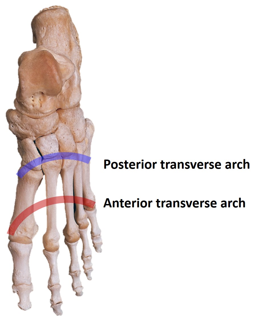
Transverse Arch of the Foot. This image shows the bony framework of the foot with the anterior and posterior components of the transverse arch highlighted. The anterior transverse arch is formed by the heads of the metatarsal bones, while the posterior transverse arch is formed by the bases of the metatarsals, cuboid, and cuneiform bones. These arches contribute to weight distribution, shock absorption, and stability during gait.
Contributed by H Chauhan, MD
(Click Image to Enlarge)
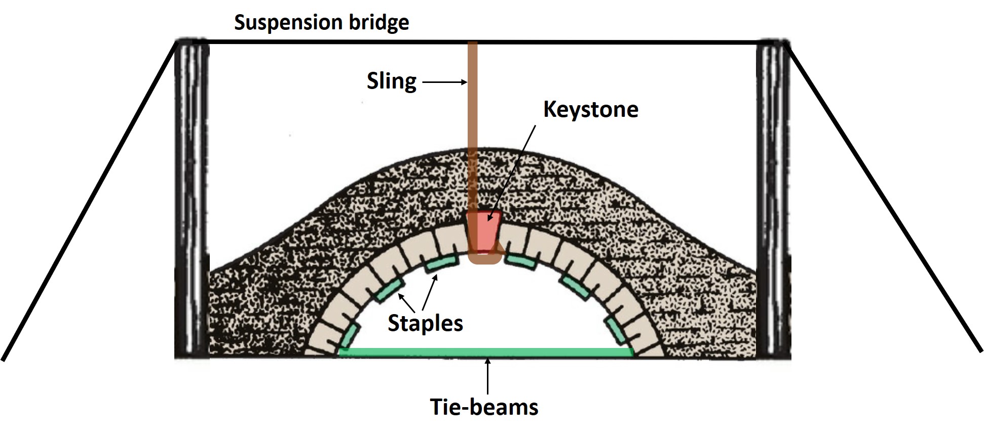
Stone Bridge Arch as an Analogy for Foot Arches. This illustration demonstrates the structural elements that maintain the stability of a stone bridge arch, including the suspension bridge, sling, keystone, staples, and tie-beams. The image is included as an analogy to illustrate the role of corresponding anatomical structures in maintaining the arches of the foot.
Snell Anatomy Plates, edited by H Chauhan, MD
(Click Image to Enlarge)
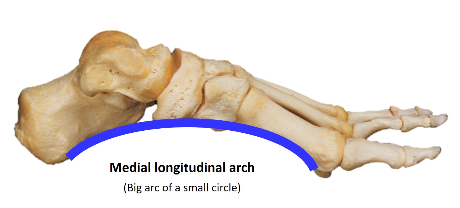
Medial Longitudinal Arch of the Foot. The medial longitudinal arch forms the primary structural curve of the foot, often described as the "big arc of a small circle." This arch consists of the medial portion of the calcaneus, talus, navicular, the 3 cuneiform bones, and the 1st to 3rd metatarsals.
Contributed by H Chauhan, MD
(Click Image to Enlarge)
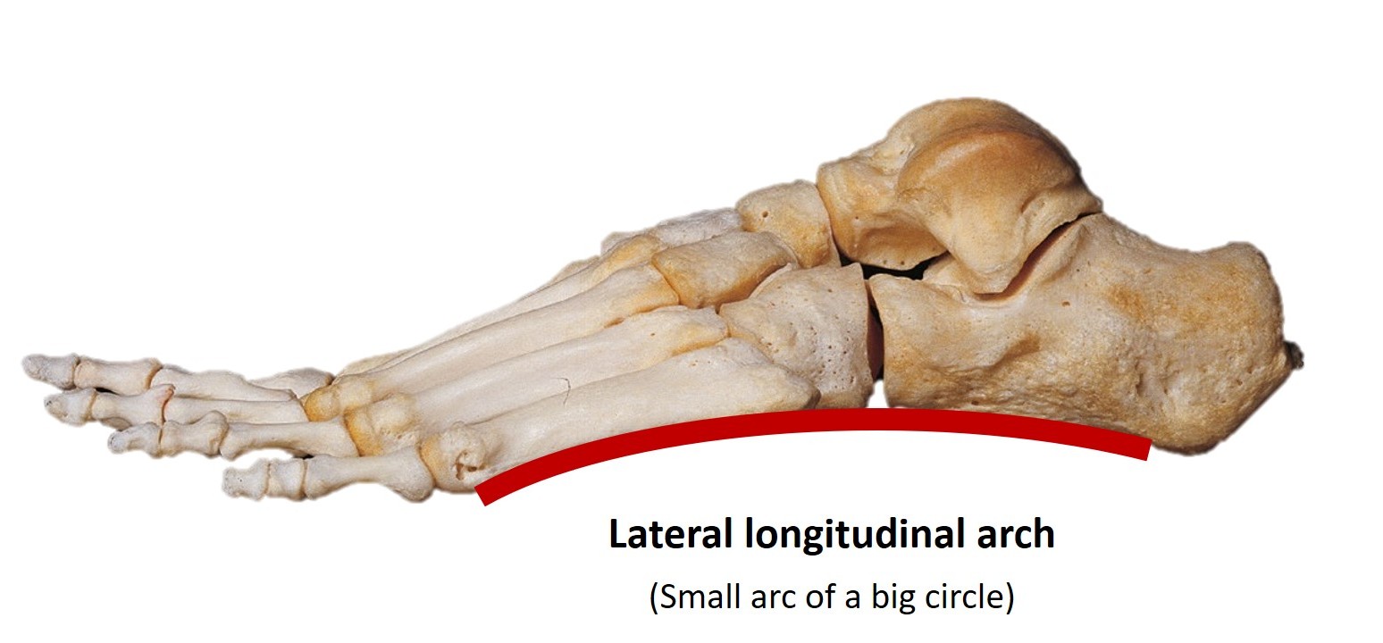
Lateral Longitudinal Arch of the Foot. The lateral longitudinal arch forms a structural curve of the foot, referred to as the "small arc of a big circle." This arch consists of the lateral portion of the calcaneus, the cuboid, and the 4th and 5th metatarsals. The lateral longitudinal arch provides stability and serves as a supportive base during weight bearing.
Contributed by H Chauhan, MD
(Click Image to Enlarge)
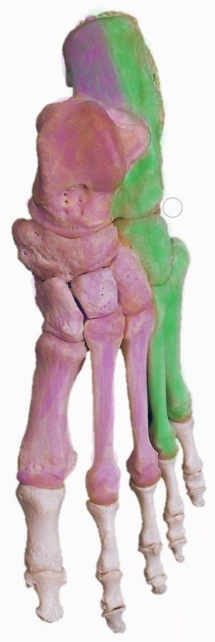
Bones Forming the Foot Arches. The medial longitudinal arch (pink) is formed by the medial portion of the calcaneus, talus, navicular, the 3 cuneiform bones, and the 1st to 3rd metatarsals. The lateral longitudinal arch (green) is formed by the lateral portion of the calcaneus, cuboid, and the 4th and 5th metatarsals.
Contributed by H Chauhan, MD
References
Babu D, Bordoni B. Anatomy, Bony Pelvis and Lower Limb: Medial Longitudinal Arch of the Foot. StatPearls. 2025 Jan:(): [PubMed PMID: 32965960]
Gwani AS, Asari MA, Mohd Ismail ZI. How the three arches of the foot intercorrelate. Folia morphologica. 2017:76(4):682-688. doi: 10.5603/FM.a2017.0049. Epub 2017 May 29 [PubMed PMID: 28553850]
Ghanem I, Massaad A, Assi A, Rizkallah M, Bizdikian AJ, El Abiad R, Seringe R, Mosca V, Wicart P. Understanding the foot's functional anatomy in physiological and pathological conditions: the calcaneopedal unit concept. Journal of children's orthopaedics. 2019 Apr 1:13(2):134-146. doi: 10.1302/1863-2548.13.180022. Epub [PubMed PMID: 30996737]
Level 3 (low-level) evidenceAsghar A, Naaz S. The transverse arch in the human feet: A narrative review of its evolution, anatomy, biomechanics and clinical implications. Morphologie : bulletin de l'Association des anatomistes. 2022 Dec:106(355):225-234. doi: 10.1016/j.morpho.2021.07.005. Epub 2021 Aug 19 [PubMed PMID: 34419345]
Level 3 (low-level) evidenceVenkadesan M, Yawar A, Eng CM, Dias MA, Singh DK, Tommasini SM, Haims AH, Bandi MM, Mandre S. Stiffness of the human foot and evolution of the transverse arch. Nature. 2020 Mar:579(7797):97-100. doi: 10.1038/s41586-020-2053-y. Epub 2020 Feb 26 [PubMed PMID: 32103182]
Sawah A, Kasture S, Bond A, Fisher L, Fisher A, Philpott M, Mason L, Molloy A. Anatomical Description of the Spring Ligament Articular Facet. Foot & ankle orthopaedics. 2024 Jul:9(3):24730114241270207. doi: 10.1177/24730114241270207. Epub 2024 Aug 26 [PubMed PMID: 39193450]
Card RK, Bordoni B. Anatomy, Bony Pelvis and Lower Limb, Foot Muscles. StatPearls. 2025 Jan:(): [PubMed PMID: 30969527]
Mostafa E, Graefe SB, Varacallo MA. Anatomy, Bony Pelvis and Lower Limb: Leg Posterior Compartment. StatPearls. 2025 Jan:(): [PubMed PMID: 30726025]
Khan IA, Mahabadi N, D’Abarno A, Varacallo MA. Anatomy, Bony Pelvis and Lower Limb: Leg Lateral Compartment. StatPearls. 2025 Jan:(): [PubMed PMID: 30137811]
Kanatli U, Yetkin H, Bolukbasi S. Evaluation of the transverse metatarsal arch of the foot with gait analysis. Archives of orthopaedic and trauma surgery. 2003 May:123(4):148-50 [PubMed PMID: 12734711]
Level 2 (mid-level) evidenceZhang L, Zhang Q, Zhong Y, Hortobagyi T, Gu Y. Effect of forefoot transverse arch stiffness on foot biomechanical response--based on finite element method. Frontiers in bioengineering and biotechnology. 2024:12():1387768. doi: 10.3389/fbioe.2024.1387768. Epub 2024 Jul 8 [PubMed PMID: 39040495]
Anderson BW, Ekblad J, Black AC, Bordoni B. Anatomy, Appendicular Skeleton. StatPearls. 2025 Jan:(): [PubMed PMID: 30571018]
Marín-Llera JC, Garciadiego-Cázares D, Chimal-Monroy J. Understanding the Cellular and Molecular Mechanisms That Control Early Cell Fate Decisions During Appendicular Skeletogenesis. Frontiers in genetics. 2019:10():977. doi: 10.3389/fgene.2019.00977. Epub 2019 Oct 11 [PubMed PMID: 31681419]
Level 3 (low-level) evidenceKlaassen Z, Shoja MM, Tubbs RS, Loukas M. Supernumerary and absent limbs and digits of the lower limb: a review of the literature. Clinical anatomy (New York, N.Y.). 2011 Jul:24(5):570-5. doi: 10.1002/ca.21102. Epub 2011 Jan 3 [PubMed PMID: 21647959]
Talamillo A, Bastida MF, Fernandez-Teran M, Ros MA. The developing limb and the control of the number of digits. Clinical genetics. 2005 Feb:67(2):143-53 [PubMed PMID: 15679824]
Balasankar G, Luximon A, Al-Jumaily A. Current conservative management and classification of club foot: A review. Journal of pediatric rehabilitation medicine. 2016 Nov 30:9(4):257-264 [PubMed PMID: 27935562]
Raj MA, Tafti D, Kiel J. Pes Planus. StatPearls. 2025 Jan:(): [PubMed PMID: 28613553]
Kopaczyńska A, Bober A, Puk A, Chwałczyńska A. Evaluation of Foot Structure in Preschool Children Based on Body Mass. Medical science monitor : international medical journal of experimental and clinical research. 2024 Apr 25:30():e943765. doi: 10.12659/MSM.943765. Epub 2024 Apr 25 [PubMed PMID: 38659197]
Guillot C, Smith T. Anatomy, Bony Pelvis and Lower Limb: Foot Arteries. StatPearls. 2025 Jan:(): [PubMed PMID: 32809747]
Takahashi LA, França GJ, Valle CED, Ferreira LRC. Assessment of the pedal arteries with Duplex Scanning. Jornal vascular brasileiro. 2020 Nov 11:19():e20200068. doi: 10.1590/1677-5449.200068. Epub 2020 Nov 11 [PubMed PMID: 34211519]
Lezak B, Varacallo MA. Anatomy, Bony Pelvis and Lower Limb, Foot Veins. StatPearls. 2025 Jan:(): [PubMed PMID: 31194435]
Karip B, Ertaş A. Abnormal vein patterns on the feet: two case reports. Folia morphologica. 2023:82(3):732-734. doi: 10.5603/FM.a2022.0053. Epub 2022 May 24 [PubMed PMID: 35607869]
Level 3 (low-level) evidenceShinaoka A, Koshimune S, Suami H, Yamada K, Kumagishi K, Boyages J, Kimata Y, Ohtsuka A. Lower-Limb Lymphatic Drainage Pathways and Lymph Nodes: A CT Lymphangiography Cadaver Study. Radiology. 2020 Jan:294(1):223-229. doi: 10.1148/radiol.2019191169. Epub 2019 Nov 19 [PubMed PMID: 31746690]
Suami H. Lymphosome concept: Anatomical study of the lymphatic system. Journal of surgical oncology. 2017 Jan:115(1):13-17. doi: 10.1002/jso.24332. Epub 2016 Jun 22 [PubMed PMID: 27334241]
Ficke J, Byerly DW. Anatomy, Bony Pelvis and Lower Limb: Foot. StatPearls. 2025 Jan:(): [PubMed PMID: 31536304]
Tang A, Bordoni B. Anatomy, Bony Pelvis and Lower Limb, Foot Nerves. StatPearls. 2025 Jan:(): [PubMed PMID: 30725977]
Lezak B, Massel DH, Varacallo MA. Peroneal Nerve Injury. StatPearls. 2025 Jan:(): [PubMed PMID: 31751049]
Kiel J, Kaiser K. Tarsal Tunnel Syndrome. StatPearls. 2025 Jan:(): [PubMed PMID: 30020645]
Desai SS, Cohen-Levy WB. Anatomy, Bony Pelvis and Lower Limb: Tibial Nerve. StatPearls. 2025 Jan:(): [PubMed PMID: 30725713]
Chang CH, Yang WT, Wu CP, Chang LW. Would foot arch development in children characterize a body maturation process? A prospective longitudinal study. Biomedical journal. 2022 Oct:45(5):828-837. doi: 10.1016/j.bj.2021.10.012. Epub 2021 Nov 2 [PubMed PMID: 34737119]
Farzadi M, Safaeepour Z, Nabavi H, Cham MB, Mousavi ME. Effect of Different Placement of Heel Rockers on Lower-Limb Joint Biomechanics in Healthy Individuals. Journal of the American Podiatric Medical Association. 2018 May:108(3):231-235. doi: 10.7547/16-052. Epub [PubMed PMID: 29932758]
Umberger BR. Stance and swing phase costs in human walking. Journal of the Royal Society, Interface. 2010 Sep 6:7(50):1329-40. doi: 10.1098/rsif.2010.0084. Epub 2010 Mar 31 [PubMed PMID: 20356877]
Geng X, Yang P, Wang X, Geng Y, Han Y. [Recognition of walking stance phase and swing phase based on moving window]. Sheng wu yi xue gong cheng xue za zhi = Journal of biomedical engineering = Shengwu yixue gongchengxue zazhi. 2014 Apr:31(2):273-8 [PubMed PMID: 25039126]
Brockett CL, Chapman GJ. Biomechanics of the ankle. Orthopaedics and trauma. 2016 Jun:30(3):232-238 [PubMed PMID: 27594929]
Leardini A, Stagni R, O'Connor JJ. Mobility of the subtalar joint in the intact ankle complex. Journal of biomechanics. 2001 Jun:34(6):805-9 [PubMed PMID: 11470119]
Welte L, Kelly LA, Lichtwark GA, Rainbow MJ. Influence of the windlass mechanism on arch-spring mechanics during dynamic foot arch deformation. Journal of the Royal Society, Interface. 2018 Aug:15(145):. doi: 10.1098/rsif.2018.0270. Epub [PubMed PMID: 30111662]
Blackwood CB, Yuen TJ, Sangeorzan BJ, Ledoux WR. The midtarsal joint locking mechanism. Foot & ankle international. 2005 Dec:26(12):1074-80 [PubMed PMID: 16390642]
Isikan UE. The values of talonavicular angles in patients with pes planus. The Journal of foot and ankle surgery : official publication of the American College of Foot and Ankle Surgeons. 1993 Sep-Oct:32(5):514-6 [PubMed PMID: 8252011]
Patil V, Shah M, Krishnani K, Jhala N. The Impact of Pes Planus on Knee Function and Its Association with Medial Compartment Knee Osteoarthritis. Journal of orthopaedic case reports. 2024 Oct:14(10):275-281. doi: 10.13107/jocr.2024.v14.i10.4888. Epub [PubMed PMID: 39381308]
Level 3 (low-level) evidenceKarataş L, Utkan Karasu A. Association of medial longitudinal arch height and stiffness with lower extremity alignment, pain, and disease severity in knee osteoarthritis: A cross-sectional study. Archives of rheumatology. 2024 Dec:39(4):641-651. doi: 10.46497/ArchRheumatol.2024.10858. Epub 2024 Dec 12 [PubMed PMID: 40060129]
Level 2 (mid-level) evidenceSeaman TJ, Ball TA. Pes Cavus. StatPearls. 2025 Jan:(): [PubMed PMID: 32310476]
Rhim HC, Kwon J, Park J, Borg-Stein J, Tenforde AS. A Systematic Review of Systematic Reviews on the Epidemiology, Evaluation, and Treatment of Plantar Fasciitis. Life (Basel, Switzerland). 2021 Nov 24:11(12):. doi: 10.3390/life11121287. Epub 2021 Nov 24 [PubMed PMID: 34947818]
Level 1 (high-level) evidenceBuchanan BK, Sina RE, Kushner D. Plantar Fasciitis. StatPearls. 2025 Jan:(): [PubMed PMID: 28613727]
Elabd K, Basudan L, Alomari MA, Almairi A. Plantar Fasciitis as a Potential Early Indicator of Elevated Cardiovascular Disease Risk. Cureus. 2024 Jun:16(6):e62007. doi: 10.7759/cureus.62007. Epub 2024 Jun 9 [PubMed PMID: 38983990]
Allam AE, Chang KV. Plantar Heel Pain. StatPearls. 2025 Jan:(): [PubMed PMID: 29763043]
RITCHIE GW, KEIM HA. A RADIOGRAPHIC ANALYSIS OF MAJOR FOOT DEFORMITIES. Canadian Medical Association journal. 1964 Oct 17:91(16):840-4 [PubMed PMID: 14217246]
Barnes CJ, Dydyk AM. Talipes Equinovarus. StatPearls. 2025 Jan:(): [PubMed PMID: 32491773]
Rieger MA, Dobbs MB. Clubfoot. Clinics in podiatric medicine and surgery. 2022 Jan:39(1):1-14. doi: 10.1016/j.cpm.2021.08.006. Epub [PubMed PMID: 34809788]
Barrie A, Varacallo MA. Clubfoot. StatPearls. 2025 Jan:(): [PubMed PMID: 31855401]
Mustari MN, Faruk M, Bausat A, Fikry A. Congenital talipes equinovarus: A literature review. Annals of medicine and surgery (2012). 2022 Sep:81():104394. doi: 10.1016/j.amsu.2022.104394. Epub 2022 Aug 18 [PubMed PMID: 36147065]
Park CH, Chang MC. Forefoot disorders and conservative treatment. Yeungnam University journal of medicine. 2019 May:36(2):92-98. doi: 10.12701/yujm.2019.00185. Epub 2019 May 14 [PubMed PMID: 31620619]
Abousayed M, Kwon JY. Hallux claw toe. Foot and ankle clinics. 2014 Mar:19(1):59-63. doi: 10.1016/j.fcl.2013.11.001. Epub 2013 Dec 19 [PubMed PMID: 24548509]
Wu DY, Lam KF. Osteodesis for hallux valgus correction: is it effective? Clinical orthopaedics and related research. 2015 Jan:473(1):328-36. doi: 10.1007/s11999-014-3938-6. Epub 2014 Oct 28 [PubMed PMID: 25349035]
Level 2 (mid-level) evidenceRay JJ, Friedmann AJ, Hanselman AE, Vaida J, Dayton PD, Hatch DJ, Smith B, Santrock RD. Hallux Valgus. Foot & ankle orthopaedics. 2019 Apr:4(2):2473011419838500. doi: 10.1177/2473011419838500. Epub 2019 May 7 [PubMed PMID: 35097321]
Kuhn J, Alvi F. Hallux Valgus. StatPearls. 2025 Jan:(): [PubMed PMID: 31971732]
Rampal V, Giuliano F. Forefoot malformations, deformities and other congenital defects in children. Orthopaedics & traumatology, surgery & research : OTSR. 2020 Feb:106(1S):S115-S123. doi: 10.1016/j.otsr.2019.03.021. Epub 2019 Oct 21 [PubMed PMID: 31648997]
Kihara T, Kimura T, Suzuki N, Hattori A, Saito M, Kubota M. Dysfunction of the Windlass Mechanism Is Associated with Hallux Rigidus: A Case-Control Study. The Journal of bone and joint surgery. American volume. 2025 Mar 19:107(6):558-564. doi: 10.2106/JBJS.24.00437. Epub 2025 Jan 31 [PubMed PMID: 39888978]
Level 2 (mid-level) evidenceDiDomenico L, Baze E, Gatalyak N. Revisiting the tailor's bunion and adductovarus deformity of the fifth digit. Clinics in podiatric medicine and surgery. 2013 Jul:30(3):397-422. doi: 10.1016/j.cpm.2013.04.004. Epub [PubMed PMID: 23827493]
Mazoteras-Pardo V, Becerro-de-Bengoa-Vallejo R, Losa-Iglesias M, Palomo-López P, López-López D, Calvo-Lobo C, Romero-Morales C, Casado-Hernández I. Degree of Impact of Tailor's Bunion on Quality of Life: A Case-Control Study. International journal of environmental research and public health. 2021 Jan 16:18(2):. doi: 10.3390/ijerph18020736. Epub 2021 Jan 16 [PubMed PMID: 33467061]
Level 2 (mid-level) evidenceSaunier J, Chapurlat R. Stress fracture in athletes. Joint bone spine. 2018 May:85(3):307-310. doi: 10.1016/j.jbspin.2017.04.013. Epub 2017 May 13 [PubMed PMID: 28512006]
Koo AY, Tolson DR. March Fracture (Metatarsal Stress Fractures)(Archived). StatPearls. 2025 Jan:(): [PubMed PMID: 30335322]