Introduction
Acute ankle sprains are among the most common musculoskeletal injuries, particularly in physically active individuals.[1] These injuries occur when excessive inversion or eversion forces exceed the tensile strength of the supporting ligamentous structures, resulting in stretching, partial tearing, or complete ligament rupture.[2] Most ankle sprains involve the lateral ligament complex, especially the anterior talofibular ligament, the weakest and most commonly injured structure. More severe injuries may also involve the calcaneofibular ligament and, rarely, the posterior talofibular ligament. Medial sprains, involving the deltoid ligament, and syndesmotic (high ankle) sprains, involving the distal tibiofibular ligaments, are less frequent but are often associated with greater morbidity. Acute sprains trigger an inflammatory response, leading to pain, swelling, and joint instability.[3]
Intrinsic risk factors include poor proprioception, a history of previous ankle sprains, ligamentous laxity, and lower extremity malalignment.[4] Extrinsic factors involve inadequate warm-up, improper footwear, participation in high-risk sports, and environmental hazards such as uneven surfaces. Athletes engaged in activities requiring rapid directional changes or jumping—such as basketball, soccer, and volleyball—are particularly susceptible.[5] Evidence-based treatment follows the POLICE principle (protection, optimal loading, ice, compression, elevation). Early functional rehabilitation is preferred over prolonged immobilization, as it promotes ligamentous healing and restores joint stability.
In moderate to severe cases, short-term bracing or taping may provide additional support. Progressive weight-bearing and neuromuscular training, including balance exercises, are essential in preventing recurrent sprains.[6] Although nonsteroidal anti-inflammatory drugs are used for pain relief, caution is advised against long-term use. Surgical intervention is reserved for cases of chronic instability or failed conservative treatment. Preventative measures, including proprioceptive training and strength conditioning, significantly reduce the risk of recurrence.
Etiology
Register For Free And Read The Full Article
 Search engine and full access to all medical articles
- 10 free questions in your specialty
- Free CME/CE Activities
- Free daily question in your email
- Save favorite articles to your dashboard
- Emails offering discounts
Learn more about a Subscription to StatPearls Point-of-Care
Etiology
Acute ankle sprains occur when external forces overwhelm the stabilizing ligamentous structures of the ankle, resulting in partial or complete tears.[7] Forced inversion is the most common mechanism and primarily affects the lateral ligament complex, particularly the anterior talofibular ligament. Less commonly, eversion injuries result in medial sprains, while dorsiflexion and external rotation forces can cause syndesmotic (high ankle) sprains. Intrinsic factors such as age, sex, body composition, proprioceptive deficits, prior sprains, and lower extremity alignment contribute to risk.[2][8] Extrinsic factors include improper footwear, training errors, and surface irregularities. Understanding these risk factors allows for targeted prevention strategies.[6]
Epidemiology
In the United States (US), more than 2 million acute ankle sprains are treated annually.[9] The incidence ranges from 2 to 7 sprains per 1000 individuals per year in the general population, with significantly higher rates among athletes.[10] Ankle sprains account for a substantial proportion of athletic injuries, particularly in sports that require cutting, jumping, or sudden changes in direction.[11] Young adults, particularly those aged 15 to 35, experience the highest incidence, and men have higher injury rates in competitive sports. Women, however, demonstrate increased susceptibility due to neuromuscular and biomechanical differences.[2][12][13]
Recurrent sprains and chronic instability are common, with up to 40% of individuals reporting persistent symptoms. Work-related ankle sprains are also significant, particularly in occupations that require prolonged standing or navigating uneven surfaces.[14] Prior literature has estimated that foot and ankle injuries account for nearly 10% of the total workplace injuries, and in 2017, over 92,000 foot and ankle injuries resulted in lost work days.[14] An increased incidence has been observed in the mining, utilities, construction, and transportation industries.[15] In the US active-duty military population, the incidence rate was 58.3 injuries per 1000 individuals per year.[16]
Pathophysiology
Most ankle sprains occur via an inversion mechanism (foot turning inward) that stresses the lateral ligament complex of the ankle.[17][18] The lateral ligament complex of the ankle consists of the anterior talofibular ligament (ATFL), calcaneofibular ligament (CFL), and posterior talofibular ligament (PTFL). These ligaments are typically injured in sequence, with the ATFL being the weakest and most commonly torn. Approximately 70% of lateral ankle sprains involve injury to the ATFL, often in isolation. The CFL is injured less frequently, typically with a dorsiflexed or inverted foot, and the PTFL is rarely torn, except in the most severe injuries, such as dislocations. By contrast, eversion injuries that damage the medial deltoid ligament are uncommon. The deltoid complex is very strong, so isolated medial ligament sprains are rare and usually occur only with significant trauma, often alongside fractures.[18]
A distinct subset of ankle sprains involves the distal tibiofibular syndesmoses, the ligaments that bind the tibia and fibula above the ankle. Syndesmotic injuries, commonly referred to as high ankle sprains, typically result from an external rotation and/or hyperdorsiflexion mechanism, rather than from isolated inversion. High ankle sprains are far less common than typical lateral sprains (roughly 1% to 10% of all ankle sprains), but they are seen more frequently in collision sports and can signify a more severe injury.
Given the force required to disrupt the syndesmosis, these injuries occur primarily in competitive athletes and may indicate instability of the ankle mortise. Severe ankle sprains can also be associated with osteochondral injuries of the talus, tears of the peroneal tendons, or other soft tissue damage that may complicate recovery. Understanding the mechanism and structures involved in an ankle sprain is crucial for guiding evaluation and management, as different injury patterns (lateral, medial, and syndesmotic) have distinct implications for treatment.
History and Physical
A comprehensive history and physical examination are crucial for accurately diagnosing acute ankle sprains, determining their severity, and ruling out associated injuries, such as fractures or syndesmotic damage. Early and accurate assessment ensures appropriate management, preventing chronic instability and long-term functional impairment.[19]
History
The clinical assessment begins with a detailed history focusing on the mechanism of injury, which provides insight into the structures involved.[20]Patients often report a twisting injury with immediate pain and swelling; a “pop” or tearing sensation may be noted in higher-grade sprains. Inversion injuries, the most common mechanism, suggest lateral ligament involvement, particularly the ATFL. Eversion injuries raise concern for medial ligament damage, while dorsiflexion-external rotation mechanisms may indicate a syndesmotic (high ankle) sprain.
Patients should describe the onset of pain, its location, and any associated symptoms, such as swelling, bruising, or instability. Weight-bearing status immediately after the injury is crucial, as the inability to bear weight may indicate a more severe sprain, fracture, or other structural injury. Additionally, prior history of ankle injuries, including recurrent sprains or chronic instability, should be elicited, as they increase the risk of long-term dysfunction.
Physical Examination
The physical examination begins with inspection, assessing for swelling, ecchymosis, and deformity. Acute ankle sprains typically present with swelling and ecchymosis localized around the injured ligaments, classically swelling over the lateral aspect of the ankle in an inversion sprain. More diffuse swelling or deformity might suggest a fracture or a high ankle sprain. Diffuse swelling suggests ligamentous injury, while localized swelling over the malleoli may indicate an occult fracture.
Palpation is performed systematically, focusing on the lateral ligaments (ATFL, CFL, PTFL), medial deltoid ligament, and the syndesmotic ligaments.[1] Tenderness along the lateral or medial malleolus necessitates evaluation for possible fracture using the Ottawa ankle rules (see below). The foot should also be palpated for associated injuries, particularly over the base of the fifth metatarsal, the navicular, and the midfoot to rule out a potential Lisfranc injury.
Systematic physical exam
- Palpation of bony landmarks
- Gently palpate the medial and lateral malleoli, base of the fifth metatarsal, and navicular bone.
- Point tenderness at these sites may indicate an occult fracture and should prompt imaging per the Ottawa ankle rules.
- Palpate proximally along the fibula up to the knee; tenderness at the proximal fibula suggests a possible Maisonneuve fracture (a spiral fibular fracture associated with syndesmosis disruption).
- Palpation of ligaments
- Examine the lateral ligaments: ATFL at the anterior aspect of the fibula, CFL just below the fibula, and PTFL posteriorly, assessing for tenderness or gaps that could indicate a rupture.
- Palpate the medial deltoid ligament for tenderness.
- Marked tenderness over the anterior-inferior tibiofibular interval (just above the ankle joint) raises concern for a syndesmotic injury.
- Range of motion and functional testing
- Assess active and passive range of motion; pain at end-range inversion is common after lateral sprains.
- Passive inversion stress may reproduce lateral ligament pain, whereas eversion stress implicates medial structures.
- Strength testing evaluates the peroneal and tibialis posterior muscles, which are critical for dynamic stabilization.
- Note any locking, catching, or crepitus, which could suggest an osteochondral fragment.
Special Tests to Assess Ligamentous Integrity
Special tests help determine the presence and extent of ligamentous injury. These maneuvers should be performed carefully, as acute pain and muscle guarding may limit accuracy immediately after injury.
- Anterior drawer test
- This is performed with the patient’s foot relaxed and positioned in approximately 20° of plantarflexion.
- Stabilize the distal tibia with one hand and draw the calcaneus and talus forward with the other.[21]
- Excessive forward translation of the talus (>8–10 mm, or >1 cm compared to the contralateral side) or a soft end-point indicates laxity or rupture of the ATFL.
- This test is most specific for ATFL injuries but may yield false negatives acutely due to pain and muscle spasm.
- Talar tilt test
- Stabilize the distal leg and invert the heel while keeping the ankle in a neutral to slight dorsiflexion position.
- Increased inversion of the talus relative to the opposite ankle (typically >15° difference) suggests injury to the CFL, often accompanied by ATFL involvement.
- Pain and guarding immediately after injury can limit reliability, as with the anterior drawer.
- Squeeze test
- Compress the tibia and fibula firmly with the patient seated at the mid-calf, then release quickly.
- Pain at the distal tibiofibular syndesmosis elicited by this maneuver raises concern for a high ankle sprain.
- External rotation stress test
- Stabilize the leg and dorsiflex the ankle, then externally rotate the foot.
- During this maneuver, pain at the anterolateral ankle or syndesmosis indicates possible syndesmotic injury.
- In some cases, this test may be performed under fluoroscopy or with stress radiographs to detect widening of the ankle mortise.
In summary, lateral ligament sprains typically exhibit maximal tenderness over the ATFL/CFL, swelling and bruising in the lateral gutter, and laxity on inversion or anterior drawer testing, if examined within a few days after injury, once acute pain has subsided. Medial ligament sprains exhibit deltoid tenderness and eversion pain, whereas syndesmotic injuries cause pain above the ankle, positive squeeze and external rotation tests, and often difficulty with weight-bearing. Always assess neurovascular status and skin integrity as well. Any gross instability, deformity, or neurovascular compromise should raise concern for fracture-dislocation rather than a simple sprain.
Grading of Ankle Sprains
Clinically, acute ankle sprains are graded by severity to guide management and prognosis.
Grade I (mild)
- This involves a slight stretching of the ligament with microscopic fiber tearing. The patient exhibits mild tenderness, minimal swelling, and no mechanical instability. Typically, weight-bearing is possible with only mild pain.
Grade II (moderate)
- This involves an incomplete ligament tear. There is moderate pain, swelling, and bruising with tenderness over the involved structures. The patient may have some difficulty with weight-bearing. On exam, the ligament is somewhat lax compared to the opposite ankle, but a definite endpoint to stress testing is still present.
Grade III (severe)
- This involves a complete rupture of 1 or more ligaments. Pain and swelling are significant, often with diffuse edema and ecchymosis, and the patient typically cannot bear weight initially. On exam, instability is evident: stress tests show gross laxity with empty end feel. Grade III injuries often involve the ATFL and CFL being torn and can be associated with additional injuries such as syndesmotic tearing or small avulsion fractures.
Accurate grading may occasionally be possible only in retrospect, since acute pain and swelling can restrict the initial examination. Despite these challenges, grading remains important for guiding treatment decisions. Grades I and II are typically managed conservatively and tend to recover more quickly, while grade III injuries often require extended protection, rehabilitation, and possibly surgery.
Evaluation
Imaging is used to distinguish ankle sprains from fractures or other injuries, and to assess severity in certain cases.
Radiograph
Standard radiographs are the first-line imaging modality when indicated.[22] The decision to order radiographs is guided by the Ottawa ankle rules, a widely validated clinical decision-making tool. According to the Ottawa criteria, radiographs should be obtained if there is bony pain in the malleolar or midfoot area, plus any of the following findings:
- Bone tenderness at the posterior edge or tip of the lateral or medial malleolus for ankle injuries
- Bone tenderness at the base of the fifth metatarsal or the navicular bone for midfoot injuries
- Inability to bear weight both immediately after the injury and in the emergency department/clinic, defined as inability to take at least 4 steps
The use of the Ottawa ankle rules yields a near 100% sensitivity for detecting clinically significant fractures, reducing the need for unnecessary radiographs by approximately 30% (see Image. Acute Ankle Sprain, Radiograph). When obtained, plain radiographs should include anteroposterior, lateral, and mortise views of the ankle.[23] Weight-bearing views are preferred if the patient can tolerate them, as they can reveal subtle joint space widening or alignment issues that nonweight-bearing films might miss. In an acute lateral ankle sprain, radiographs are often normal or may reveal only soft tissue swelling.
However, imaging is essential to detect fractures that can mimic or accompany sprains, such as malleolar fractures, fractures of the base of the fifth metatarsal, or navicular fractures. Small avulsion fragments may also be seen, typically arising from the fibula at the anterior talofibular ligament attachment or from the talus at the deltoid ligament attachment in more severe (grade III) sprains (see Images. Grade III Acute Ankle Sprain, Talar Tilt Test, Radiograph and Grade III Acute Ankle Sprain, Anterior Drawer Test, Radiograph). These fragments usually heal with conservative treatment unless they are significantly displaced.
If a high ankle sprain is suspected, stress radiographs can be useful. An external rotation or gravity stress view of the ankle may demonstrate widening of the distal tibiofibular or medial clear space, indicating syndesmotic ligament disruption. A medial clear space greater than 4 mm or a tibiofibular clear space greater than 6 mm on stress imaging suggests syndesmotic instability. Significant widening of stress views usually warrants surgical stabilization (syndesmotic screw or fixation, discussed later).
Magnetic Resonance Imaging
Magnetic resonance imaging (MRI) is the most sensitive imaging modality for assessing soft tissue injuries, including ligamentous tears, syndesmotic injury, osteochondral defects, and tendon involvement. MRI is indicated when:
- Pain persists beyond 6 weeks despite appropriate conservative treatment
- High ankle sprain is suspected
- There are concerns for osteochondral injuries or occult fractures
MRI is advantageous for assessing soft tissue injuries in the ankle region and is typically reserved for planning primary ligamentous surgical repair. Diagnosis criteria of acute ligamentous injury include morphologic and signal intensity changes within and around the suspected ligament. Healthy ligaments of the ankle complex appear as thin, linear, low-signal-intensity structures when uninjured.
With an acute injury, intrasubstance edema can be seen on fat-suppressed or T2-weighted imaging as increased signal intensity within the ligament. In contrast, chronic injury to the ligament is characterized by thickening, elongation, and irregular contouring of the suspected ligament with no significant soft tissue edema, marrow changes, or hemorrhaging noted by increased signal intensity. The ATFL, CFL, and PTFL are best viewed on coronal and axial views with the foot placed in dorsiflexion, and a cross-sectional thickness of 3 mm or less is recommended.
Ultrasound
Ultrasound offers a dynamic and cost-effective method for evaluating ligament integrity, identifying joint effusions, and detecting peroneal tendon subluxation. An ultrasound's ability to assess ligament laxity in real-time makes it particularly useful, although its accuracy depends heavily on operator expertise. Ultrasound may serve as an alternative when MRI is not available or can complement the clinical examination.
Computed tomography
Computed tomography scans provide a detailed evaluation of complex fractures, particularly in cases of suspected osteochondral injuries or when standard radiographs are inconclusive. They are rarely needed for routine ankle sprains but may be useful in evaluating subtle fractures or syndesmotic injuries.
Treatment / Management
Nonoperative Management
Most acute ankle sprains, including virtually all grade I and II and most grade III sprains, are managed nonoperatively.[24] The goal of treatment is to allow ligament healing while maintaining the ankle's range of motion, strength, and stability. Strong evidence supports functional rehabilitation over extended immobilization for treating sprains. Key components of conservative management include:(B2)
Protection and rest
- During the acute phase (the first 1–3 days), protect the ankle from further injury and refrain from engaging in activities that cause pain. For more severe sprains, a short period of immobilization may be helpful. For example, a grade III sprain may benefit from up to 10 days of immobilization in a cast or walking boot with crutches (nonweight-bearing), especially if the patient cannot walk due to pain.
- However, extended immobilization beyond this initial period is discouraged. Clinical guidelines recommend against casting for more than 4 weeks in even the most severe sprains, as prolonged immobilization can lead to weakness and stiffness without improving long-term outcomes. Lighter protection, like a semi-rigid brace or splint, and early motion are preferred as soon as it is safe.
Ice and compression
- Aggressive cold therapy using ice or cold packs and compression with an elastic bandage or sleeve should be initiated immediately after the injury. These measures help limit swelling, reduce pain, and may expedite recovery. Cryotherapy in the acute stage has a high level of evidence supporting its use in improving short-term outcomes. Elevating the injured ankle above heart level, especially in the first 48 hours, also helps reduce swelling. The acronym RICE often summarizes this acute care approach: rest, ice, compression, elevation.
Early controlled motion
- Early range-of-motion exercises should be initiated after the initial pain and swelling begin to subside, often within the first 3–7 days, depending on severity. Rather than immobilizing the ankle, functional treatment encourages gentle motion as tolerated. For example, guided exercises such as ankle circles, writing the alphabet with the toes, and gentle calf stretches can be introduced in the early phase. Early mobilization has been shown to accelerate recovery; patients who initiate functional rehabilitation early can return to work or sports more quickly than those who remain immobilized for prolonged periods.
Bracing and support
- During the rehabilitation process, a support device is often recommended. A semi-rigid ankle brace, air stirrup, or ankle taping can provide external support to protect the ligaments from re-injury during healing. For mild sprains, a simple compression wrap or soft brace may be sufficient; for moderate to severe sprains, a more supportive brace or boot is useful during the initial couple of weeks.
- After the acute phase, a functional ankle brace that limits inversion/eversion but allows plantar/dorsiflexion is typically worn during rehab and high-risk activities. This not only stabilizes the joint while ligaments heal, but also helps restore proprioceptive feedback. Study results indicate that functional bracing or taping for 4–6 weeks after a sprain is more effective than prolonged casting in preventing recurrent injury.
Physical therapy and rehabilitation
- A structured rehabilitation program is critical for optimal recovery, especially for grade II and III sprains. Rehabilitation typically progresses in phases. Phase 1 (acute/early) focuses on reducing pain and swelling (RICE, nonsteroidal anti-inflammatory drugs [NSAIDs], and protected weight-bearing) while restoring range of motion through gentle exercises. Phase 2 (subacute) involves the addition of strengthening exercises, particularly for the peroneal muscles and calf, once swelling has diminished and range of motion is improving.
- Resistance band exercises, such as eversion, toe/heel raises, and stationary cycling, are commonly introduced. Phase 3 (advanced) emphasizes proprioception and functional training, incorporating balance exercises (such as single-leg stands and wobble board training), agility drills, and sport-specific movements to retrain neuromuscular control. Maintaining cardiovascular fitness and strength in the rest of the body is encouraged throughout all phases. Supervised physical therapy can be very helpful in the early stages to ensure proper exercise technique and progress. However, some study results show similar long-term outcomes with a home exercise program once the patient is educated.
Medications
- Ankle sprains can be painful, especially in the first week. NSAIDs, such as ibuprofen or naproxen, are commonly used to reduce pain and inflammation in the acute setting. Short courses of NSAIDs can help patients mobilize earlier by managing pain. Some recent evidence suggests that simple analgesics like acetaminophen (paracetamol) or even short-term use of opioids can be as effective as NSAIDs for pain control in acute sprains. These alternatives may be considered in patients who cannot take NSAIDs. Regardless, pain management should be balanced with safety—for most ankle sprains, strong pain medications are only needed for a few days at most.
- Follow-up should ensure that the patient’s pain, swelling, and function are improving. Suppose a patient fails to progress or has persistent ankle instability (giving-way sensation) despite proper rehab. In that case, they should be re-evaluated for possible underlying issues (such as a missed syndesmotic injury or an osteochondral lesion). These refractory cases may require advanced imaging or orthopedic referral, as they could be candidates for surgical intervention if conservative measures have truly failed.[25] (A1)
Indications for Surgical Management
Fortunately, acute surgical intervention is rarely required for ankle sprains.[26] The default approach is nonoperative treatment, and as noted above, this is successful for most injuries. However, certain situations warrant orthopedic consultation and consideration of surgery. Operative management may be indicated in the following scenarios:
Chronic ankle instability
- The most common indication for lateral ligament surgery is not an acute sprain, but persistent instability after repeated sprains. Patients who continue to have ankle giving-way episodes, pain, or inability to resume activities despite extensive nonoperative management, typically 3–6 months of bracing and therapy, are candidates for surgical stabilization. Essentially, a grade III sprain or even lesser sprains that do not heal sufficiently and result in chronic lateral ankle instability may require repair of the ligaments to restore stability.[27]
Acute high ankle injury with instability
-
If an ankle sprain involves the syndesmosis and results in diastasis (separation) of the distal tibia and fibula seen on imaging, surgical stabilization of the ankle mortise is indicated. Radiographic signs include widening the medial clear space or obvious instability on stress views. These injuries frequently occur alongside fractures—such as a high fibular fracture seen in a Maisonneuve injury—but even in isolated ligamentous syndesmotic ruptures, operative fixation with screws or suture-button devices is typically necessary to prevent long-term ankle instability.
Associated fractures or osteochondral lesions
-
In some severe sprains, a fragment of bone may be avulsed—such as a piece from the distal fibula or tibia at the ligament attachment site—or a loose fragment of cartilage or bone may form within the joint. When these avulsion fragments are large and significantly displaced, or when an osteochondral fragment is inside the joint, surgical intervention is warranted to fix or remove the fragment. These injuries are more complex than a simple sprain and often require treatment similar to fractures, necessitating reduction and fixation.
High-performance athletes with grade III tears
- In elite athletes or individuals with high-demand occupations, primary surgical repair of a completely torn ligament may be considered to expedite return to play. Evidence on this is mixed, and routine acute surgery is not recommended for the general population due to longer recovery time and risk of stiffness. However, some sports medicine surgeons perform an acute Broström repair of the ATFL/CFL in a professional athlete with a confirmed complete rupture. This aims to allow a more stable and faster rehab for return to sport. Thus, individualized consideration is given in elite athletes or in cases where timing is critical, balancing the benefits against the risks.
Medial (deltoid) or combined injuries
- Isolated deltoid ligament sprains are usually treated nonoperatively. But if the deltoid ligament is completely ruptured in an unstable bimalleolar injury (medial clear space widening) or if chronic medial instability develops, surgical repair or reconstruction of the deltoid may be indicated. This is more often seen in fracture scenarios than pure sprains.
Notably, acute surgical repair of lateral ankle ligaments is generally not recommended for first-time sprains. Meta-analyses indicate that conservative treatment typically yields outcomes equal to or superior to those of immediate surgery, and it avoids surgical risks such as infection, nerve injury, and joint stiffness. As a result, surgery is reserved for the specific scenarios described earlier. Clinical practice guidelines give a very low grade of recommendation for acute surgical intervention in routine cases. The widely accepted approach is to pursue thorough nonoperative treatment first and consider surgery only if significant instability or persistent pain remains.
Surgical Techniques and Outcomes
The specific procedure is tailored to the injury pattern when surgery is indicated.
Lateral ligament repair (Broström procedure)
- For chronic lateral ankle instability that persists despite conservative treatment, the gold-standard surgical option is an anatomic repair of the torn ATFL and, if necessary, the CFL.[28] The classic procedure is the Broström repair, which is often reinforced with the Gould modification, involving the suturing of the inferior extensor retinaculum to the fibula to strengthen the reconstruction.
- Suture anchors may be used in the fibula to secure the ligaments. This anatomic repair has become the standard procedure for treating chronic lateral ankle instability. Multiple studies' results have demonstrated good to excellent results in roughly 90% of patients after a Broström repair, with restored stability and high rates of return to sports.
- The Broström-Gould technique preserves normal ankle kinematics by reconstructing the original ligaments in their anatomical positions. Patients are generally immobilized for 2 weeks after repair, then start range of motion exercises and progress to strengthening. A brace is worn for support during the first 2 to 3 months of return to activity. Most athletes can return to play within approximately 3 to 4 months postoperatively with appropriate rehabilitation. (A1)
Ligament reconstruction (tendon grafts)
- In some cases, such as long-standing instability with poor-quality native ligaments, failed prior repairs, or generalized ligamentous laxity, a nonanatomic reconstruction is performed instead of or in addition to a primary repair. Numerous procedures exist (eg, Chrisman-Snook, Watson-Jones, Evans procedures), typically involving the use of a tendon graft, often the peroneus brevis tendon, to reconstruct the lateral ligament complex.
- These reconstructions substitute the torn ligaments with tendon tissue routed through bone tunnels in the fibula and talus/calcaneus. While they can successfully stabilize the ankle, they are nonanatomic and may restrict some motion; subtalar stiffness is a known possible complication. Now, tendon reconstructions are generally reserved for revision cases or when the Broström repair is not feasible. An anatomic Broström repair is the preferred initial surgical approach for most patients with isolated chronic ankle instability.[29]
Syndesmosis fixation
- Surgical stabilization of the syndesmosis is required in cases of high ankle sprain with diastasis. This typically involves placement of 1 or 2 cortical screws across the distal tibiofibular joint to hold the tibia and fibula in proper alignment while the syndesmotic ligaments heal. An alternative device is a suture-button implant (flexible fixation), which may allow physiologic micro-motion. These devices maintain the distal tibiofibular approximation without the need for a rigid screw. In either case, the goal is to restore the normal ankle mortise; failure can lead to chronic pain and arthritis.
- Syndesmotic screws are often removed 3 to 4 months later, whereas suture-button implants are usually left in place. Postoperatively, the patient is kept nonweight-bearing for several weeks to protect the repair, followed by gradual weight-bearing in a boot.
Medial ligament repair
- In the uncommon situation where deltoid ligament surgery is needed, typically performed with ankle fracture fixation or for chronic medial instability, the superficial and deep deltoid ligaments may be repaired to the medial malleolus or reconstructed using a tendon graft if required. This procedure is rarely performed in isolation and is usually part of a broader surgical strategy for complex ankle injuries.
Arthroscopy and management of associated injuries
- Ankle arthroscopy is frequently used as an adjunct during the surgical management of ankle sprains, especially in chronic cases. Arthroscopy enables direct visualization of the joint, allowing for the identification of any osteochondral lesions, loose bodies, or hypertrophic scar tissue that may be causing impingement. For example, in chronic sprains, patients can develop anterolateral impingement due to scar tissue in the lateral gutter or osteochondral defects in the talus. These can be debrided or fixed arthroscopically during the ligament surgery.
- Arthroscopy can also confirm reduction of the ankle mortise during syndesmosis fixation. In cases of impingement syndromes or unexplained post-sprain pain, arthroscopy with debridement can alleviate symptoms and is sometimes performed even without a formal ligament repair if instability is not a concern.[29]
With appropriate patient selection and technique, surgical outcomes for ankle sprains are generally very good. Lateral ligament repair using the Broström procedure yields excellent stability and function in approximately 90% of patients, with a low rate of complications. In most cases, patients typically report improved stability, fewer or no episodes of giving way, and the ability to return to sports at a preinjury level. Performing surgery unnecessarily or too early in a patient who might have improved with rehabilitation can result in risks that outweigh the benefits. These risks include infection, nerve irritation, sural or superficial peroneal nerve injury, wound complications, and ankle stiffness.
Therefore, the current best practice is to reserve surgery for clear cases of persistent mechanical instability or confirmed significant injury that cannot heal on its own. Even after surgical stabilization, rehabilitation remains essential. Postoperative therapy focusing on range of motion and peroneal muscle strengthening is necessary to achieve the best outcome. Early mobilization protocols following Broström repair have demonstrated improved functional scores, although this must be balanced against the risk of overstretching the repair.
Differential Diagnosis
When evaluating an acute ankle injury, clinicians must consider a broad differential diagnosis beyond the classic lateral ankle sprain (see Table. Differential Diagnosis of Acute Ankle Sprains). Lateral ankle ligament injuries, particularly involving the ATFL, CFL, and PTFL, are the most common and typically result from inversion injuries. However, medial ankle sprains involving the deltoid ligament occur with eversion mechanisms and present with medial pain and swelling. Syndesmotic sprains, or high ankle sprains, result from external rotation or hyperdorsiflexion and cause pain above the ankle joint, often with difficulty bearing weight.
Fractures, including ankle and Lisfranc injuries, should be considered when there is significant pain, deformity, or inability to bear weight. Osteochondral lesions, talar dome osteochondritis dissecans, and ankle impingement syndromes may present with persistent or mechanical symptoms beyond the typical healing course. Soft tissue pathologies such as peroneal tendon injuries, posterior tibial tendon dysfunction, Achilles tendon rupture, and extensor/flexor tendon injuries should be evaluated through specific physical exam maneuvers and imaging. Finally, chronic or atypical symptoms may point to tarsal coalition or nerve entrapment syndromes, requiring careful clinical assessment and often advanced diagnostic imaging. A thorough differential helps prevent missed diagnoses and guides targeted management.
Table. Differential Diagnosis of Acute Ankle Sprains
|
Lateral ankle ligament injuries (ATFL, CFL, PTFL) |
Lateral pain, swelling, bruising after inversion injury; positive anterior drawer/talar tilt tests | Represents the most common true acute ankle sprain; see below for other differentials |
| Medial ankle sprains (deltoid ligament injury) | Medial pain, swelling, bruising after eversion injury; tenderness over the deltoid ligament | Less common; the mechanism is usually eversion or significant trauma |
| Syndesmotic (high ankle) sprains | Pain above ankle, worse with external rotation; positive squeeze/external rotation tests; difficulty weight-bearing | Tenderness over the syndesmosis (anterior distal tibiofibular joint); often more severe; the mechanism is external rotation or dorsiflexion |
| Ankle fractures | Inability to bear weight; bony tenderness; deformity or crepitus; abnormal x-rays | Fracture evident on radiographs; more severe pain/swelling, possible deformity |
| Osteochondral lesions of the talus | Persistent deep ankle pain, catching or locking, swelling; history of trauma | Symptoms often persist after a sprain should resolve; MRI/CT shows a talar defect |
| Peroneal tendon injuries (tendinopathy, subluxation, dislocation) | Lateral ankle pain, swelling, tenderness behind the lateral malleolus; possible snapping sensation | Pain/posterior to lateral malleolus; pain with resisted eversion; subluxation visible on dynamic exam |
| Achilles tendon rupture | Sudden pain and "pop" in posterior ankle/calf; palpable gap; positive Thompson test | Weak/absent plantarflexion; cannot stand on tiptoes; swelling posterior, not lateral or medial |
| Posterior tibial tendon dysfunction | Medial ankle/foot pain, swelling, arch collapse, difficulty with inversion | Progressive flatfoot deformity; weakness of inversion; not acute traumatic |
| Talar dome osteochondritis dissecans | Chronic ankle pain, swelling, occasional catching/locking | Symptoms outlast typical sprain recovery; imaging shows a subchondral bone lesion |
| Ankle impingement syndrome (anterior/posterior) | Chronic pain with forced dorsiflexion (anterior) or plantarflexion (posterior); may develop after trauma | Pain with specific ankle movement; bony impingement on imaging, not acute injury |
| Tarsal coalition | Chronic hindfoot pain, recurrent sprains, and rigid flatfoot in adolescents | Decreased subtalar motion; radiographs/CT reveal coalition |
| Lisfranc injury | Midfoot pain/swelling, plantar ecchymosis, pain with forefoot abduction/pronation | Pain localized to midfoot, not ankle; instability with midfoot stress; weight-bearing x-ray shows diastasis |
| Extensor or flexor tendon injuries | Pain, swelling, weakness in toe extension/flexion; possible palpable defect | Weakness/loss of active toe movement; localized tenderness along the tendon |
| Nerve entrapment syndromes (sural, tarsal tunnel) | Burning/tingling, numbness in nerve distribution; worsened with activity | Neuropathic symptoms; Tinel sign positive over affected nerve; not traumatic |
| Septic or inflammatory arthritis | Diffuse swelling, erythema, warmth, severe pain; often febrile, systemic symptoms | Joint aspiration shows infection or crystals; nontraumatic, often bilateral if inflammatory |
ATFL, anterior talofibular ligament; CFL, calcaneofibular ligament; CT, computed tomography; MRI, magnetic resonance imaging; PTFL, posterior talofibular ligament
Prognosis
The prognosis following an acute ankle sprain depends on the severity of the injury, adherence to rehabilitation, and presence of predisposing factors such as recurrent instability or inadequate neuromuscular control. While most patients recover fully with conservative management, a subset may develop chronic symptoms or recurrent sprains. A history of ankle sprain represents a significant risk factor for future injuries, underscoring the importance of recurrence prevention as a central component of effective management.
Athletes may benefit from using an ankle brace or tape during high-risk sports for 6–12 months after the injury to prevent further recurrence. Follow-up should ensure that the patient’s pain, swelling, and function are improving. Suppose a patient does not improve or continues to experience ankle instability, characterized by a giving-way sensation, despite appropriate rehabilitation. In that case, they should be re-evaluated for possible underlying issues, such as an unrecognized syndesmotic injury or an osteochondral lesion. These refractory cases may require advanced imaging or orthopedic referral, as they could be candidates for surgical intervention if conservative measures have truly failed.
Short-Term Prognosis
Patients with grade I sprains typically recover within 1–2 weeks with early mobilization and functional rehabilitation. These injuries involve mild ligament stretching without significant structural damage, allowing for rapid return to daily activities and sports participation. Grade II sprains, characterized by partial ligament tears, require 3–6 weeks for recovery, with progressive weight-bearing and strength training essential for restoring stability. Grade III sprains, involving complete ligament rupture, have a more variable prognosis, often necessitating 6–12 weeks of rehabilitation, and in some cases, surgical intervention for persistent instability.
Long-Term Outcomes and Complications
While most individuals recover fully, up to 40% of patients experience residual symptoms, including:
- Chronic ankle instability: Recurrent sprains due to insufficient ligament healing, neuromuscular deficits, or proprioceptive dysfunction
- Chronic ankle instability significantly increases the risk of long-term joint degeneration.
- Persistent pain and swelling: Often due to unrecognized intra-articular injuries such as osteochondral lesions, peroneal tendon pathology, or impingement syndromes
- Posttraumatic osteoarthritis
- Although uncommon after a single sprain, repeated injuries and inadequate rehabilitation can lead to degenerative joint changes, particularly in athletes and individuals with high-impact occupational demands.
Factors Influencing Prognosis
- Consistent early rehabilitation: This includes balance training and strength exercises, significantly improving outcomes and lowering the risk of reinjury.
- Biomechanical and neuromuscular deficiencies, including poor proprioception, muscle imbalances, and inadequate postural control, increase the likelihood of reinjury.
- Severity and location of injury: High ankle (syndesmotic) sprains often require prolonged recovery due to the complexity of the ligamentous structures involved.
- Previous history of ankle sprains: Individuals with a prior history are more likely to experience chronic instability and may require extended rehabilitation or surgical intervention.
Return-to-Sport Timeline
- Grade I: 1–2 weeks
- Grade II: 3–6 weeks
- Grade III: 6–12 weeks or longer (with possible surgical consideration)
Complications
The complications with acute ankle sprains that can manifest include the following:
- Chronic instability: There can be a persistent feeling of the ankle "giving way" due to insufficient ligament healing or inadequate rehabilitation.
- Recurrent sprains: There can be an increased susceptibility to future sprains due to inadequate treatment or incomplete recovery.
- Osteoarthritis: The development of joint degeneration can occur over time due to altered biomechanics or injury to cartilage.
- Tendon injuries: Damage to the Achilles tendon or peroneal tendons can occur due to compensatory movements or overuse during recovery.
- Nerve damage: Potential injury to the superficial peroneal nerve can lead to sensory deficits or pain.
- Ligamentous laxity: Stretching or tearing of ligaments can lead to prolonged instability and necessitate surgical intervention.
- Ankle fractures: In severe cases, an ankle sprain may be accompanied by a fracture of the fibula, tibia, or other bones.
- Posttraumatic osteochondral lesions: Damage to the articular cartilage often results in long-term pain and functional limitations.
- Deep vein thrombosis: Prolonged immobility or swelling following injury can lead to blood clots.
- Chronic swelling and pain: There is a risk of persistent edema and discomfort due to incomplete resolution of soft tissue injury.
- Impaired range of motion: Restricted movement can be caused by ligament tightness, muscle weakness, or joint stiffness.
Deterrence and Patient Education
Education for patients who sustain an acute ankle sprain is crucial in promoting proper healing and preventing long-term complications. Patients should be instructed on the importance of following the POLICE principle (protection, optimal loading, ice, compression, and elevation) during the initial phase of recovery to manage swelling and pain. Early mobilization and gradual weight-bearing are emphasized to prevent stiffness and maintain joint function. Patients should be educated on recognizing signs of complications such as persistent swelling or instability, which may require further imaging or intervention. Emphasis on a structured rehabilitation program, including strengthening and proprioception exercises, is essential to reduce the risk of recurrent sprains and long-term instability.
Enhancing Healthcare Team Outcomes
Interprofessional communication and care coordination are vital in enhancing patient outcomes following an acute ankle sprain. Collaboration among healthcare professionals, including primary care clinicians, orthopedic surgeons, physical therapists, and athletic trainers, ensures comprehensive care tailored to the patient's specific needs. Clear communication among these professionals allows for timely diagnosis, effective treatment plans, and seamless transitions between care stages. For example, a primary care clinician may initiate an acute management plan, while a physical therapist provides rehabilitation strategies to restore strength and proprioception.
Regular updates and shared information between healthcare professionals help track progress, adjust treatment protocols, and address complications promptly. This coordinated approach minimizes the risk of reinjury, improves recovery time, and optimizes functional outcomes. Moreover, patient education consistently delivered by various professionals promotes adherence to rehabilitation exercises, improves compliance, and reduces the likelihood of chronic instability or recurring sprains. Ultimately, cohesive interprofessional care promotes more effective recovery trajectories and enhances long-term patient well-being.[30][31]
Media
(Click Image to Enlarge)
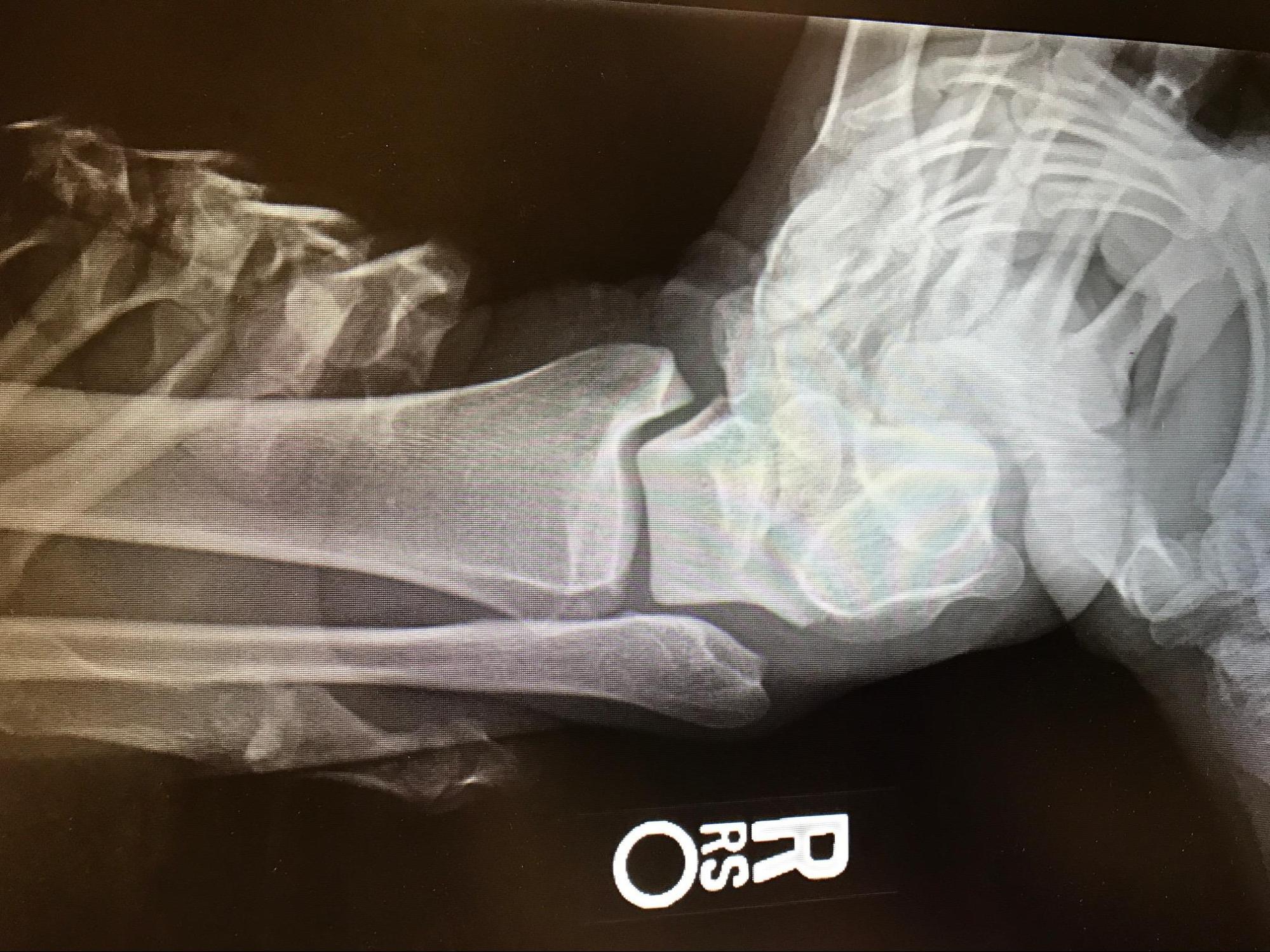
Grade III Acute Ankle Sprain, Talar Tilt Test, Radiograph. This image shows a grade III ankle sprain, demonstrated by the talar tilt test stress radiograph. Note the varus positioning of the talus on the tibia.
Contributed by MA Dreyer, DPM, FACFAS
(Click Image to Enlarge)
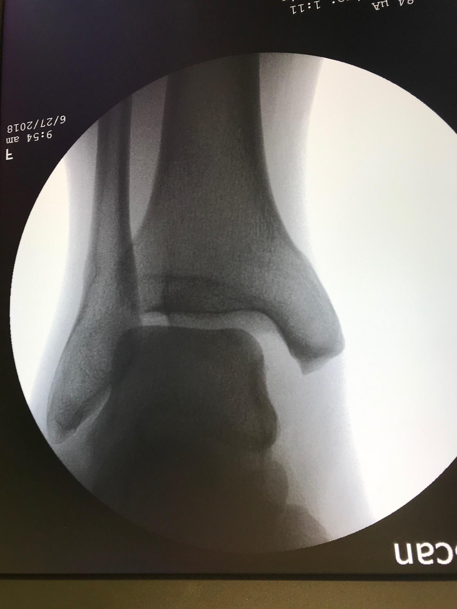
Acute Ankle Sprain, Radiograph. This image demonstrates deltoid and syndesmotic insufficiency in the setting of an acute ankle sprain, evidenced by medial gutter widening.
Contributed by MA Dreyer, DPM, FACFAS
(Click Image to Enlarge)
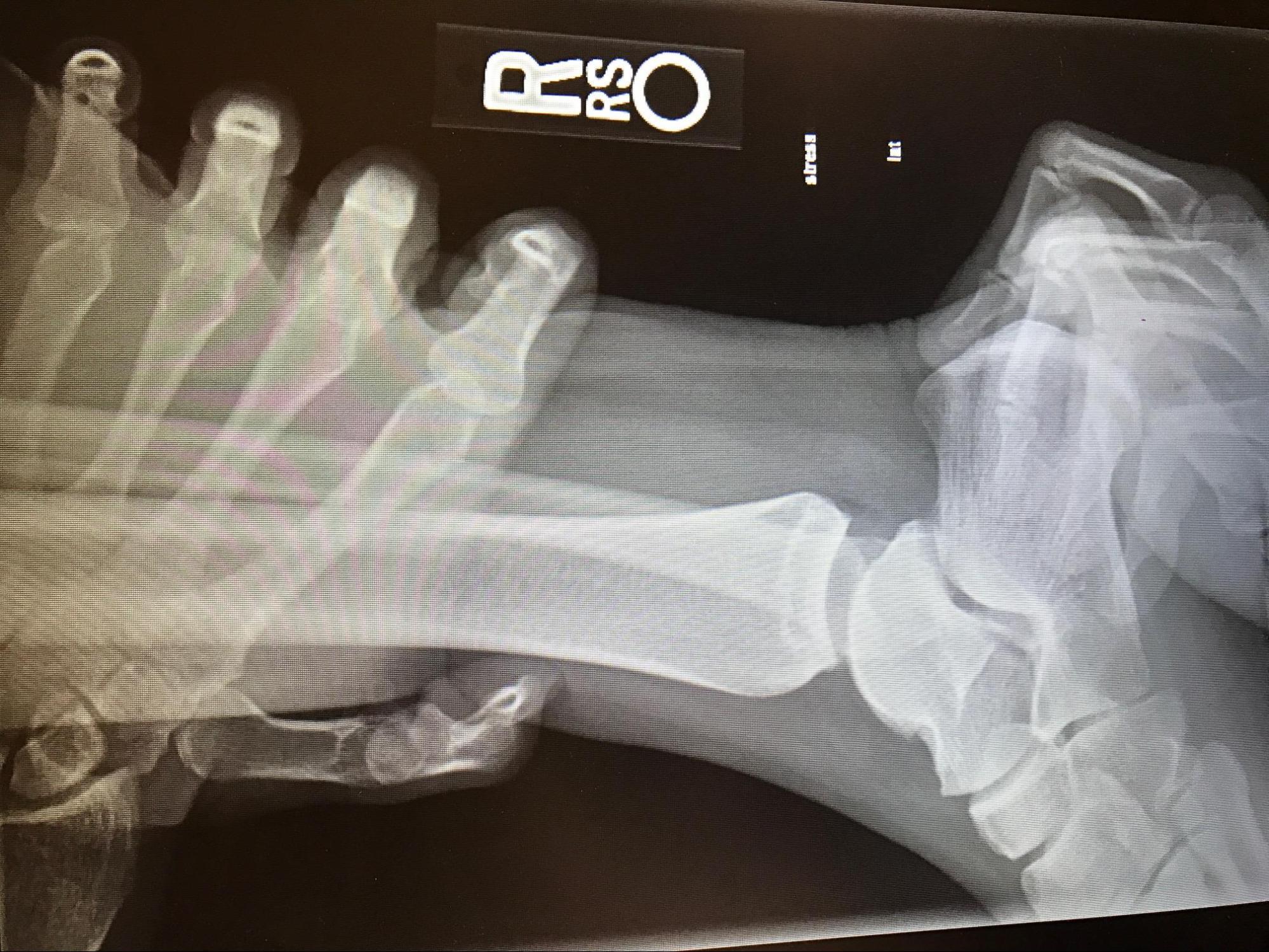
Grade III Acute Ankle Sprain, Anterior Drawer Test, Radiograph. This image shows an ankle sprain demonstrated by an anterior drawer stress radiograph. Note the anterior translation of the talus on the tibia.
Contributed by MA Dreyer, DPM, FACFAS
References
Hertel J. Functional Anatomy, Pathomechanics, and Pathophysiology of Lateral Ankle Instability. Journal of athletic training. 2002 Dec:37(4):364-375 [PubMed PMID: 12937557]
Doherty C, Delahunt E, Caulfield B, Hertel J, Ryan J, Bleakley C. The incidence and prevalence of ankle sprain injury: a systematic review and meta-analysis of prospective epidemiological studies. Sports medicine (Auckland, N.Z.). 2014 Jan:44(1):123-40. doi: 10.1007/s40279-013-0102-5. Epub [PubMed PMID: 24105612]
Level 1 (high-level) evidenceTyler TF, McHugh MP, Mirabella MR, Mullaney MJ, Nicholas SJ. Risk factors for noncontact ankle sprains in high school football players: the role of previous ankle sprains and body mass index. The American journal of sports medicine. 2006 Mar:34(3):471-5 [PubMed PMID: 16260467]
Trojian TH, McKeag DB. Single leg balance test to identify risk of ankle sprains. British journal of sports medicine. 2006 Jul:40(7):610-3; discussion 613 [PubMed PMID: 16687483]
Doherty C, Bleakley C, Hertel J, Caulfield B, Ryan J, Delahunt E. Recovery From a First-Time Lateral Ankle Sprain and the Predictors of Chronic Ankle Instability: A Prospective Cohort Analysis. The American journal of sports medicine. 2016 Apr:44(4):995-1003. doi: 10.1177/0363546516628870. Epub 2016 Feb 24 [PubMed PMID: 26912285]
Bahr R, Krosshaug T. Understanding injury mechanisms: a key component of preventing injuries in sport. British journal of sports medicine. 2005 Jun:39(6):324-9 [PubMed PMID: 15911600]
Level 3 (low-level) evidenceDelahunt E, Remus A. Risk Factors for Lateral Ankle Sprains and Chronic Ankle Instability. Journal of athletic training. 2019 Jun:54(6):611-616. doi: 10.4085/1062-6050-44-18. Epub 2019 Jun 4 [PubMed PMID: 31161942]
Mason J, Kniewasser C, Hollander K, Zech A. Intrinsic Risk Factors for Ankle Sprain Differ Between Male and Female Athletes: A Systematic Review and Meta-Analysis. Sports medicine - open. 2022 Nov 18:8(1):139. doi: 10.1186/s40798-022-00530-y. Epub 2022 Nov 18 [PubMed PMID: 36399159]
Level 1 (high-level) evidenceWaterman BR, Owens BD, Davey S, Zacchilli MA, Belmont PJ Jr. The epidemiology of ankle sprains in the United States. The Journal of bone and joint surgery. American volume. 2010 Oct 6:92(13):2279-84. doi: 10.2106/JBJS.I.01537. Epub [PubMed PMID: 20926721]
Level 2 (mid-level) evidenceHerzog MM, Kerr ZY, Marshall SW, Wikstrom EA. Epidemiology of Ankle Sprains and Chronic Ankle Instability. Journal of athletic training. 2019 Jun:54(6):603-610. doi: 10.4085/1062-6050-447-17. Epub 2019 May 28 [PubMed PMID: 31135209]
Ferran NA, Maffulli N. Epidemiology of sprains of the lateral ankle ligament complex. Foot and ankle clinics. 2006 Sep:11(3):659-62 [PubMed PMID: 16971255]
Gribble PA,Bleakley CM,Caulfield BM,Docherty CL,Fourchet F,Fong DT,Hertel J,Hiller CE,Kaminski TW,McKeon PO,Refshauge KM,Verhagen EA,Vicenzino BT,Wikstrom EA,Delahunt E, Evidence review for the 2016 International Ankle Consortium consensus statement on the prevalence, impact and long-term consequences of lateral ankle sprains. British journal of sports medicine. 2016 Dec; [PubMed PMID: 27259753]
Level 3 (low-level) evidenceRoos KG, Kerr ZY, Mauntel TC, Djoko A, Dompier TP, Wikstrom EA. The Epidemiology of Lateral Ligament Complex Ankle Sprains in National Collegiate Athletic Association Sports. The American journal of sports medicine. 2017 Jan:45(1):201-209. doi: 10.1177/0363546516660980. Epub 2016 Oct 1 [PubMed PMID: 27573356]
Galasso A, Caughman AM, Griffith A, Hoch C, Rex J, Scott DJ, Gross CE. A Detailed Analysis of Workplace Foot and Ankle Injuries. Foot & ankle specialist. 2024 Feb 29:():19386400241233844. doi: 10.1177/19386400241233844. Epub 2024 Feb 29 [PubMed PMID: 38424705]
Conti SF, Silverman L. Epidemiology of foot and ankle injuries in the workplace. Foot and ankle clinics. 2002 Jun:7(2):273-90 [PubMed PMID: 12462110]
Waterman BR, Belmont PJ Jr, Cameron KL, Deberardino TM, Owens BD. Epidemiology of ankle sprain at the United States Military Academy. The American journal of sports medicine. 2010 Apr:38(4):797-803. doi: 10.1177/0363546509350757. Epub 2010 Feb 9 [PubMed PMID: 20145281]
Level 2 (mid-level) evidenceCarto C, Lezak B, Varacallo MA. Anatomy, Bony Pelvis and Lower Limb: Distal Tibiofibular Joint (Tibiofibular Syndesmosis). StatPearls. 2025 Jan:(): [PubMed PMID: 31613435]
Khan IA, Varacallo MA. Anatomy, Bony Pelvis and Lower Limb, Foot Talus. StatPearls. 2025 Jan:(): [PubMed PMID: 31082130]
Maduka GC, Jakusonoka R, Maduka DC, Yusuf N. Conservative Management of Acute Lateral Ligaments of the Ankle Injuries: An Analytical Literature Review. Cureus. 2023 Oct:15(10):e47709. doi: 10.7759/cureus.47709. Epub 2023 Oct 26 [PubMed PMID: 37965420]
Zhang X, Wu J, Liang D, Ruan B, Gao Q. Effects of Different Intervention Methods on Postural Control in Athletes with Chronic Ankle Instability: A Randomized Controlled Trial. Journal of sports science & medicine. 2025 Jun:24(2):332-340. doi: 10.52082/jssm.2025.332. Epub 2025 Jun 1 [PubMed PMID: 40469867]
Level 1 (high-level) evidenceEhrlichman LK, Gonzalez TA, Macaulay AA, Ghorbanhoseini M, Kwon JY. Gravity Reduction View: A Radiographic Technique for the Evaluation and Management of Weber B Fibula Fractures. The archives of bone and joint surgery. 2017 Mar:5(2):89-95 [PubMed PMID: 28497098]
Kerkhoffs GM, Rowe BH, Assendelft WJ, Kelly K, Struijs PA, van Dijk CN. Immobilisation and functional treatment for acute lateral ankle ligament injuries in adults. The Cochrane database of systematic reviews. 2002:(3):CD003762 [PubMed PMID: 12137710]
Level 1 (high-level) evidenceBachmann LM, Kolb E, Koller MT, Steurer J, ter Riet G. Accuracy of Ottawa ankle rules to exclude fractures of the ankle and mid-foot: systematic review. BMJ (Clinical research ed.). 2003 Feb 22:326(7386):417 [PubMed PMID: 12595378]
Level 1 (high-level) evidenceFranz PB, de Souza Júnior JR, de Lima HC, de Souza Diniz E, de Brito Aguiar R, Geremia JM, Mansur H, de Cássia Marqueti R, Durigan JLQ. Acute Lateral Ankle Sprain Impairs Function and Strength Without Altering Muscle or Tendon Stiffness: A Controlled Observational Study. Orthopaedic surgery. 2025 Jul:17(7):2082-2092. doi: 10.1111/os.70082. Epub 2025 May 29 [PubMed PMID: 40442868]
Level 2 (mid-level) evidenceGrosdent S, Léonard F, Demoulin C, Aguilaniu A, Hidalgo B, Durieux N. Effectiveness of manual techniques, exercise therapy, or combined treatments in the management of ankle sprains or chronic ankle instability in adult athletes: a systematic review protocol. JBI evidence synthesis. 2025 Jul 1:23(7):1501-1509. doi: 10.11124/JBIES-24-00057. Epub 2025 Apr 4 [PubMed PMID: 40181743]
Level 1 (high-level) evidenceChaudhry H, Simunovic N, Petrisor B. Cochrane in CORR ®: surgical versus conservative treatment for acute injuries of the lateral ligament complex of the ankle in adults (review). Clinical orthopaedics and related research. 2015 Jan:473(1):17-22. doi: 10.1007/s11999-014-4018-7. Epub 2014 Oct 25 [PubMed PMID: 25344407]
Järvelä T, Weitz H, Järvelä K, Alavaikko A. A novel reconstruction technique for chronic lateral ankle instability: comparison to primary repair. International orthopaedics. 2002:26(5):314-7 [PubMed PMID: 12378362]
Kerkhoffs GM, Handoll HH, de Bie R, Rowe BH, Struijs PA. Surgical versus conservative treatment for acute injuries of the lateral ligament complex of the ankle in adults. The Cochrane database of systematic reviews. 2007 Apr 18:(2):CD000380 [PubMed PMID: 17443501]
Level 1 (high-level) evidenceKarlsson J, Eriksson BI, Bergsten T, Rudholm O, Swärd L. Comparison of two anatomic reconstructions for chronic lateral instability of the ankle joint. The American journal of sports medicine. 1997 Jan-Feb:25(1):48-53 [PubMed PMID: 9006691]
Sprouse RA, McLaughlin AM, Harris GD. Braces and Splints for Common Musculoskeletal Conditions. American family physician. 2018 Nov 15:98(10):570-576 [PubMed PMID: 30365284]
Barelds I, van den Broek AG, Huisstede BMA. Ankle Bracing is Effective for Primary and Secondary Prevention of Acute Ankle Injuries in Athletes: A Systematic Review and Meta-Analyses. Sports medicine (Auckland, N.Z.). 2018 Dec:48(12):2775-2784. doi: 10.1007/s40279-018-0993-2. Epub [PubMed PMID: 30298478]
Level 1 (high-level) evidence