Introduction
Chylothorax is the accumulation of chyle in the pleural cavity. Chyle is derived from the Greek word chylos, which means "juice." Chyle is the milky bodily fluid formed in the lacteal system of the intestine. Chylothorax can result from trauma, surgical complications, malignancies, or congenital abnormalities. This rare but serious condition was first described in the 17th century by Bartloet and has recently received special attention due to innovative management strategies yielding favorable outcomes.[1][2][3][4]
Small- and medium-chain triglycerides consumed in the diet are easily broken down into free fatty acids by intestinal enzymes and absorbed into the portal circulation. However, the large molecules of complex long-chain triglycerides cannot be broken down by the intestinal lipases. Instead, they combine with phospholipids, cholesterol, and cholesterol esters to form chylomicrons in the jejunum. These large molecules are then absorbed into the lymphatic system of the small intestine to form chyle. The lymphatic drainage from the intestine joins with that from the lower extremities to form the thoracic duct system, which ultimately drains into the systemic circulation. If the integrity of the thoracic duct is breached, the milky, lipid-rich chyle leaks into the surrounding structures.
Chylothorax occurs when chyle leaks into the pleural cavity due to damage to the thoracic duct. With an average production of approximately 2.4 L/d, a considerable amount of chyle can quickly accumulate in the pleural cavity.
The management of chylothorax in patients largely depends on the underlying cause and typically includes dietary modification and drainage of the pleural space. Definitive interventions, whether surgical or percutaneous lymphatic procedures, should be considered for patients with persistently high volumes of chylous output or prolonged leaks to prevent complications such as malnutrition.[5][4]
Etiology
Register For Free And Read The Full Article
 Search engine and full access to all medical articles
- 10 free questions in your specialty
- Free CME/CE Activities
- Free daily question in your email
- Save favorite articles to your dashboard
- Emails offering discounts
Learn more about a Subscription to StatPearls Point-of-Care
Etiology
The etiology of chylothorax can be classified into 3 broad categories—nontraumatic (spontaneous), traumatic, and idiopathic. Historically, nontraumatic chylothorax was more common, accounting for about two-thirds of cases. Recently, traumatic chylothorax, particularly postoperative chylothorax, has accounted for over 50% of cases reported in the literature.[6][7][4]
Nontraumatic Chylothorax
Nontraumatic chylothorax can arise from the following causes:
- Congenital chylothorax can occur as an isolated condition or in association with other lymphatic abnormalities, such as lymphangiectasis, lymphangiomatosis, tuberous sclerosis, congenital heart disease, or chromosomal abnormalities such as trisomy 21 or Turner syndrome.
- Neoplastic chylothorax is the most common cause of nontraumatic chylothorax. Various cancers, including lymphoma, chronic lymphocytic leukemia, lung cancer, esophageal cancer, and metastatic carcinoma, have been implicated in chylothorax. Interestingly, the incidence of chylothorax in patients with lymphoma has relatively decreased, likely due to earlier diagnosis and treatment of lymphoma, which avoids the late complication of chylothorax.
- Infectious chylothorax is primarily observed in developing countries as a complication of tuberculous lymphadenitis. Other infections that can cause chylothorax include aortitis, histoplasmosis, and filariasis.
- Rare causes of chylothorax reported in the literature include Castleman disease, sarcoidosis, Kaposi sarcoma, yellow nail syndrome, Noonan syndrome, Down syndrome, Waldenström macroglobulinemia, macroglobulinemia, amyloidosis, venous thrombosis, thoracic radiation, and goiter. All these reported conditions can lead to obstruction or destruction of the thoracic duct, resulting in chylothorax.
- Parenteral nutrition therapy is also associated with chylothorax. Rapid infusion of total parenteral nutrition (TPN) with high triglyceride concentrations can exceed the thoracic duct's capacity to drain, leading to chyle leakage into the surrounding pleural space and resulting in chylothorax.[8]
Traumatic Chylothorax
Traumatic chylothorax results from disruption of the thoracic duct at any point along its course in the mediastinum. This can occur as a complication of surgical procedures or follow blunt or penetrating chest trauma.[9]
- Postoperative chylothorax is the most common form of chylothorax in modern medicine. The risk varies depending on the type of surgery. Esophagostomy has the highest risk, with a 5% to 10% incidence of postoperative chylothorax, followed by lung resection with mediastinal lymph node dissection, which carries a 3% to 7% risk. Other procedures, such as mediastinal tumor resection, thoracic aneurysm repair, sympathectomy, and surgeries in the lower neck and mediastinum, also carry a risk of chylothorax. Ingesting milk before surgery may improve visualization of the thoracic duct in the surgical field by causing whitish discoloration, although this has not been formally studied.
- Nonsurgical posttraumatic chylothorax can occur following procedures such as central line placement, pacemaker implantation, and embolization of pulmonary arteriovenous malformations.
- Penetrating chest injuries, including gunshot and stab wounds, can directly damage the thoracic duct, leading to chylothorax.
- Blunt trauma to the chest or thoracic spine can also disrupt the thoracic duct without apparent injury to the surrounding structure, leading to chylothorax.
- Additionally, chylothorax has been reported after blasting injuries and even minor incidents such as coughing and sneezing.
Idiopathic Chylothorax
Idiopathic causes account for nearly 10% of all chylothorax cases. While esophageal resection is considered the most common iatrogenic cause, case reports have also documented idiopathic chylothorax occurring during pregnancy, triggered by labor.[10]
Epidemiology
Chylothorax, whether traumatic or nontraumatic, is a rare condition, accounting for up to 3% of all pleural effusions. Traumatic cases are more common than nontraumatic ones. Iatrogenic causes often arise from thoracic surgeries, particularly esophageal procedures, due to the proximity and variable anatomy of the thoracic duct. The incidence of iatrogenic chylothorax in esophageal resections can be as high as 4%, given the proximity of the thoracic duct to the esophagus and its variable anatomy.
Among nontraumatic causes, malignancy is the most common, accounting for nearly one-third of all chylothorax cases. Lymphoma is responsible for 70% to 75% of malignant chylothorax cases, with non-Hodgkin lymphoma being the most frequent cause. Although metastatic epithelial tumors can also lead to chylothorax, this is relatively uncommon. Other nontraumatic causes together account for about one-fifth of all cases.[11]
Pathophysiology
Anatomy of the Lymphatic System
Understanding the anatomy of the lymphatic system is crucial to understanding chylothorax. The lymphatic system comprises lymphatic vessels, lymph nodes, and the cisterna chyli. The thoracic duct is the central collecting vessel of the lymphatic system, and it drains 3 quarters of the lymph in the body into the venous bloodstream. The lymphatic system, which connects the lymphatics of various organs, is an essential network for fluid circulation throughout the body. The lymph from the lower limbs finally terminates at the para-aortic nodes. They join with the lymph from the viscera of the pelvis and form bilateral lumbar trunks. The intestinal trunks consist of large lymphatic vessels that receive lymph from the stomach, intestine, pancreas, and spleen.
The cisterna chyli is a triangular dilation of the lymphatic system located in the retroperitoneum, just posterolateral to the abdominal aorta adjacent to the second lumbar vertebra. The thoracic duct starts from the cisterna chyli and is located at the second or third lumbar vertebra level. The duct enters the thorax through the aortic hiatus of the diaphragm and ascends in the posterior mediastinum. The thoracic duct is positioned between the descending thoracic aorta on the left and the azygos vein on the right. At the fifth thoracic vertebra level, the thoracic duct inclines to the left to enter the superior mediastinum. Then, it ascends behind the aortic arch and the thoracic part of the left subclavian artery, between the left side of the esophagus and the left pleura, to the thoracic inlet.
Finally, the thoracic duct terminates by opening into the junction of the left subclavian and jugular veins. However, the duct may also open into either of the great veins near the intersection or divide into several smaller vessels before termination. In adults, the thoracic duct varies from 38 to 45 cm in length and is approximately 3 to 5 mm in diameter at its commencement. However, it narrows at the mid-thoracic level and then slightly dilates before its termination. A breach in the integrity of the thoracic duct anywhere along its course in the mediastinum will lead to the seepage of chyle into the pleural cavity, resulting in chylothorax.[11]
Composition of Chyle
Chyle comprises chylomicrons, which are aggregates of long-chain triglycerides, cholesterol esters, and phospholipids. Chyle is also rich in lymphocytes, primarily T lymphocytes, with concentrations ranging from 400 to 6800 cells. Chyle has a similar electrolyte concentration to plasma but is rich in immunoglobulins and fat-soluble vitamins.[11]
History and Physical
The clinical features of chylothorax depend on the underlying cause. Small chylothoraces can be asymptomatic and detected incidentally. Patients with large chylothoraces usually present with signs and symptoms caused by the mechanical effect of compression on lung expansion. Progressive breathlessness decreases exercise capacity. Chest pressure is a common presenting complaint, while fever and chest pain are usually absent. Patients can accumulate a large amount of chyle without any complaints if the fluid accumulation is gradual and the respiratory system acclimates. Posttraumatic chylothorax can present up to 10 days after the inciting trauma. In postsurgical patients, chylothorax is often first detected as a pleural effusion or by persistent drainage from indwelling chest tubes.
Depending on the amount and location of the fluid, decreased breath sounds and dullness to percussion may be present on physical examination. Approximately 80% of chylothorax cases are unilateral. Due to the anatomy of the thoracic duct, the right side is more commonly affected than the left, accounting for two-thirds of cases.
Evaluation
Further evaluation of chylothorax depends on the suspected cause and the availability of resources.[12]
Chest Radiograph
Chest radiographs, routinely performed to evaluate dyspnea, particularly in postoperative and traumatic patients, can detect unilateral pleural effusion. Chylothorax appears as a homogeneous density obliterating the costophrenic and cardiophrenic angles. A routine chest radiograph cannot differentiate chylothorax from other causes of pleural effusion (see Image. Chest x-Ray Showing Chylothorax With Homogeneous Density).
Thoracic Ultrasound
Ultrasound is increasingly used to evaluate patients with pleuropulmonary pathology. Similar to other effusions, chylothorax appears as an isodense echoic region without septation or loculation. However, ultrasound cannot differentiate chylothorax from other causes of pleural effusion.
Computed Tomography of the Chest
Computed tomography (CT) scan is more sensitive than chest radiographs and ultrasound for diagnosing chylothorax. Routine chest CT can show the cisterna chyli in approximately 2% of cases. Due to its high fat content, it appears as a low-attenuation tubular area in the posterior mediastinum. A CT scan can also reveal the cause of chylothorax, such as mass lesions or obstructions in the posterior mediastinum, thoracic malignancy, or evidence of trauma.
Magnetic Resonance Imaging of the Chest
Magnetic resonance imaging (MRI) of the chest shows cisterna chyli in 100% of cases and can be used to better evaluate chylothorax. However, MRI is not an optimal investigation for thoracic pathology, so it is rarely used in clinical practice.
Conventional Lymphangiography
Lymphangiography is a technique used to delineate the lymphatic system. In modern medicine, this test is rarely used due to the availability of minimally invasive alternatives that are equally precise. This technique injects a dye like methylene blue, which stains the lymphatics, into the web spaces between the toes. A longitudinal or transverse cutaneous incision at the base of the first metatarsal bone is made to expose a lymphatic vessel with blue staining after dissection of the surrounding tissue.
The isolated lymphatic vessel is then cannulated using a 30-gauge needle. After accessing the lymphatic vessel, 10 mL of lipiodol is slowly injected at 0.2 to 0.4 mL/min. Serial fluoroscopic spot images are obtained every 5 to 10 minutes as the lipiodol travels upward. If the lipiodol does not reach the area of interest, normal saline can be injected at the same rate to push the lipiodol further. This technique can reliably show any leakage in the thoracic duct leading to chylothorax.
A further modification, called nodal lymphangiogram, has recently been developed. In this procedure, a targeted lymph node is selected, and ethiodized oil is injected into the cortex of the lymph node at a rate of 1 mL/min for a total of 10 mL. Serial spot radiographs of the pelvis, abdomen, and thorax are then acquired to follow the progression of the dye. This intranodal lymphoscintigraphy is sensitive, technically more accessible, and has fewer complications. Lymphograms can be therapeutic in up to 40% of cases, as the high-density oil used in the procedure can close leaks during the procedure.
Nuclear Lymphoscintigraphy
This technique has been used more commonly than traditional lymphangiography to delineate the lymphatic system. In this procedure, Tc99m-labeled human diethylenetriaminepentaacetic acid is injected bilaterally into the subcutaneous lesions of the dorsum of the foot. Sequentially, anterior and posterior images of the chest are obtained using a gamma camera to identify the leak. This technique can be combined with integrated low-dose CT scans with single-photon emission CT (SPECT) to get more accurate SPECT/CT images. Radio nuclear localization using lymphoscintigraphy has been shown to correlate with traditional lymphangiography and surgical localization of leaks.
Laboratory Testing
Fluid analysis: Thoracentesis and fluid analysis are the diagnostic studies of choice for chylothorax. All fluid samples should be sent for white blood cell count with differential, glucose, lactic dehydrogenase, total protein level, cytology, microbiology smear, and culture. Chyle is rich in lymphocytes, accounting for 80% of all cells. The lymphocytes are predominantly a polyclonal population of T cells. If chylothorax is suspected based on the color of the fluid, additional tests such as pH, total triglyceride levels, and cholesterol levels should be performed.
Color: Based on the fat content in chylothorax, the appearance of the fluid can be milky white, serous, or serosanguineous. The absence of a milky appearance does not exclude chylothorax. After centrifugation of the pleural fluid, the supernatant is opaque in chylothorax and cholesterol effusions but clear in empyema. The milkiness is caused by suspended leukocytes and debris.
Lipid analysis: Measurement of pleural fluid triglyceride content is crucial for diagnosing chylothorax. Unlike any other body fluid, chylothorax is rich in long-chain fatty acids absorbed from the small intestine. A pleural fluid triglyceride concentration of more than 110 mg/dL typically confirms the diagnosis of chylothorax. However, 15% of chylothorax cases may have triglyceride concentrations less than 110 mg/dL, influenced by the time of the last meal and dietary fat content. If clinical suspicion remains high, lipid electrophoresis should be performed. Detection of chylomicrons in the pleural fluid via lipoprotein electrophoresis confirms chylothorax. The total cholesterol level in chylothorax is typically less than 200 mg/dL.
Other composition: The composition of the chyle is similar to plasma. Chylothorax fluid is typically alkaline, with a pH of 7.4 to 7.8.
Pseudochylothorax or Chyliform Effusions
Pseudochylothorax refers to milky-white effusions with a gross appearance similar to chylothorax, but these are less common than the classical chylothorax. These effusions are reported to be long-standing exudate pleural effusions resulting from various underlying conditions. They contain a high cholesterol concentration, which imparts a milky or white appearance. In long-standing exudative pleural effusions, cholesterol is released from cell membranes into the fluid and becomes trapped in the pleural cavity. However, unlike classical chylothorax, pseudochylothorax does not contain chylomicrons or long-chain fatty acids.
Pseudochylothorax is commonly associated with tuberculosis and chronic rheumatoid pleural effusion due to the high cell concentration. Other causes include yellow nail syndrome and paragonimiasis. In pseudochylothorax, cholesterol concentration is typically more than 200 mg/dL, triglyceride levels are less than 110 mg/dL, and the cholesterol-to-triglyceride ratio is always more than 1. Cholesterol crystals may be visible in dried pleural fluid slides under polarized light, appearing as rectangular plates with notched edges. The presence of these cholesterol crystals is virtually diagnostic of pseudochylothorax.
Treatment / Management
The management of chylothorax depends on its cause and may include interventions such as dietary modification, pleurodesis, and thoracic duct ligation. Recently, somatostatin/octreotide, midodrine, and sirolimus have been used to reduce chyle formation. New surgical techniques such as pleurovenous or pleuroperitoneal shunting and thoracic duct embolization have shown success. Most patients benefit from a staged care plan, starting with the least invasive options and progressing to more invasive techniques if necessary.[13][14][15][16]
Dietary Therapy
Decreasing or eliminating long-chain fatty acids from the diet can decrease chyle production and promote spontaneous leak closure. To effectively minimize chyle formation, a diet with less than 5 kcal of fat per meal is recommended. However, this approach may result in fat deficiency and malnutrition over time. Venous fat hemorrhage can remedy some of this therapy's shortcomings. To address these issues, small-chain and medium-chain fatty acids can be included in the diet, while long-chain fatty acids may be supplemented intravenously.
Thoracentesis
Intermittent therapeutic thoracentesis, or the use of an indwelling catheter for home drainage, is commonly used in the initial management of nontraumatic and nonsurgical traumatic effusions to alleviate dyspnea caused by pleural fluid. This technique is effective for gradual pleural fluid accumulation. For postsurgical chylothorax, a chest tube is often used. Prolonged pleural fluid drainage can lead to significant malnutrition and immunoglobulin loss, increasing infection risk. Therefore, continuous pleural fluid drainage should generally be limited to less than 2 weeks. Surgical intervention is advised if pleural fluid drainage exceeds 1.5 L daily.
Pleurodesis
Pleurodesis is indicated for patients who persistently reaccumulate fluid despite dietary modifications and repeated thoracenteses. The procedure is performed by placing a chest tube for drainage through video-assisted thoracic surgery (VATS) with talc insufflation, which can be effective in up to 80% of chylothorax cases. Additionally, concomitant ligation of the thoracic duct during surgical pleurodesis can further enhance success by preventing additional chyle formation.
Thoracic Duct Ligation
Thoracic duct ligation, performed using video-assisted thoracic surgery, is considered for patients who do not respond to dietary modifications and pleurodesis. While lymphedema is a known complication, it generally resolves over several months due to the development of collateral lymphatic-venous communications.
Thoracic Duct Embolization and Disruption
Percutaneous catheterization and embolization, involving needle disruption of the thoracic duct and cisterna chyli, have become increasingly utilized for both traumatic and nontraumatic chylothorax. The procedure begins with a pedal lymphangiogram and fluoroscopic visualization of retroperitoneal lymphatics, accessed through transabdominal needle cannulation. After cannulating the cisterna chyli, the catheter is advanced with thoracic duct installation of contrast to localize the leak. The affected thoracic segment is then embolized using coils and surgical glues.
Emerging Therapies
Somatostatin and octreotide reduce gastric, pancreatic, and biliary secretions, thereby decreasing the total flow of gastric lymphatics. Due to reduced chyle formation and flow rates, these agents can promote spontaneous closure of thoracic duct leaks. This technique is reported to be effective in many cases of spontaneous, congenital, and postoperative chylothorax, as well as chylothorax due to malignancy. However, the optimal dose and duration of treatment are not precise.
Sirolimus, used to treat lymphangiomyomatosis, is also known to decrease the incidence of chylothorax in these patients.
Shunting chylous pleural fluid into the venous system or peritoneal cavity can effectively resolve chylothorax. This can be achieved through a pleural venous or pleuroperitoneal shunt. Two types of pleuroperitoneal shunts are available: the Denver pleuroperitoneal shunt, which is an active pump requiring manual pumping, and Le Veen's pleuroperitoneal shunt, which is a passive pump. The primary advantage of shunting is the recycling of nutritionally rich chyle back into the body. Pleural venous shunting, which transfers chylous fluid from the pleural space to the subclavian or jugular vein, has been successfully performed in patients with yellow nail syndrome and postsurgical chylothorax.
Differential Diagnosis
When diagnosing chylothorax, it is crucial to consider a range of differential diagnoses that may present with similar symptoms. Various conditions can lead to pleural effusions and must be distinguished from chylothorax to ensure accurate diagnosis and appropriate treatment.
Potential differential diagnoses to consider include:
- AIDS-related complex
- Congestive heart failure
- Thoracic empyema
- Exudative pleural effusion
- Hemothorax
- Malignant and parapneumonic pleural effusion
- Pseudochylothorax
Prognosis
Chylothorax is an uncommon complication following cardiothoracic surgery, with an incidence rate of approximately 1.8%. Due to its rarity, it is challenging to conduct trials with adequate sample sizes to compare treatment options. A consensus on the optimal timing for surgical intervention does not exist after the failure of initial nonoperative management. Consequently, providing the highest standard of evidence-based recommendations for specific treatment approaches is challenging. However, given the high morbidity and mortality, prolonged hospital stays, and need for reintervention associated with chylothorax, considering early reoperation following diagnosis in postoperative cardiothoracic patients is reasonable.[17]
Most patients benefit from a staged care plan, progressing from least invasive to more invasive techniques. Overall, outcomes for patients with chylothorax from benign causes are favorable, while those with malignancy have more guarded outcomes.[18] A significant advancement in classifying chyle leaks following esophagectomy has been achieved through a Delphi Consensus by the Esophagectomy Complications Consensus Group (ECCG)—a collaborative network of 24 high-volume surgical centers across 14 countries. This classification system categorizes chyle leaks based on their response to treatment—type I requires enteral dietary modification, type II necessitates TPN, and type III demands interventional or surgical treatment. Additionally, chyle leaks are further divided by output volume—type A with less than 1 L of daily output and type B with more than 1 L of daily output.
The new ECCG consensus nomenclature and classification are expected to address gaps in future studies. Although retrospective application is not precise, it supports the approach that conservative management has been successful for type I and II leaks. This typically involves halting standard enteral nutrition in favor of TPN or enteral medium-chain triglycerides. In addition, it remains unclear whether TPN and enteral medium-chain triglycerides are equally effective or if they should be used sequentially if TPN and enteral rest achieve the desired reduction in output.
Evidence for using octreotide primarily comes from managing chyle leaks of various origins, including idiopathic and postsurgical chylothoraces. While most studies suggest that octreotide is a safe and modestly effective treatment, its specific benefit in the context of esophageal resection surgery and the relevance of leak type remain uncertain.[19]
Complications
Patients with chylothorax can face significant complications that impact their health and quality of life. A primary concern is malnutrition due to the loss of chyle, which contains essential fats, proteins, and fat-soluble vitamins. This nutritional deficit can lead to weight loss, muscle wasting, and weakened immune function.
Additionally, the ongoing loss of lymphocytes in the chyle can result in immunosuppression, increasing susceptibility to infections. Persistent pleural effusions can cause respiratory distress and impaired lung function, while prolonged chyle leakage may lead to electrolyte imbalances and fluid depletion. Effective management of these complications requires comprehensive strategies, including nutritional support, infection control, and careful monitoring of fluid and electrolyte balance.
Deterrence and Patient Education
Multiple factors can leave patients vulnerable to infections, including secondary malnutrition, underlying illnesses and their immunosuppressive therapies, the use of various devices (such as indwelling pleural catheters, tube thoracostomy, and central vascular catheters), and prolonged hospitalizations. The interprofessional healthcare team should collaboratively work to mitigate these risks to improve individual clinical outcomes. Nonetheless, deterrence and patient education are critical components in managing chylothorax effectively and preventing complications.
Patients should be educated on the importance of adhering to dietary modifications, such as a low-fat diet supplemented with medium-chain triglycerides, to help reduce chyle production and minimize effusion. They need to recognize early signs of recurrence or complications, such as increased shortness of breath or signs of infection, and seek prompt medical attention if these symptoms occur. Additionally, informing patients about the need for long-term monitoring and follow-up care can improve compliance and engagement with their treatment plan. By empowering patients with knowledge and strategies, healthcare professionals can enhance outcomes and reduce the likelihood of adverse effects related to chylothorax.
Pearls and Other Issues
Chylothorax is caused by disruption of the thoracic duct, resulting in chyle entering the pleural space. This can be seen in many conditions, of which trauma and malignancy are the leading causes. Diagnosis is based on pleural fluid analysis, which typically shows triglyceride levels greater than 110 mg/dL or the presence of chylomicrons.
Clinical pearls for managing chylothorax emphasize the importance of early diagnosis and intervention to prevent complications and improve outcomes. Key strategies involve promptly initiating dietary modifications, such as a low-fat diet supplemented with medium-chain triglycerides, to reduce chyle production. Regularly monitoring pleural effusions and effective drainage techniques are crucial for managing symptoms. Several surgical and nonsurgical approaches can be used to treat chylothorax, with somatostatin analogs currently proving effective in treatment.
Understanding the importance of a multidisciplinary approach enhances patient care, as effective coordination between pulmonologists, thoracic surgeons, dietitians, and other specialists is crucial for comprehensive treatment. Clinicians should also remain vigilant for signs of complications such as malnutrition and immunosuppression, which necessitate timely intervention to optimize patient recovery and quality of life.
Enhancing Healthcare Team Outcomes
Chylothorax has numerous causes and complex management requirements. Therefore, it is most effectively managed by an interprofessional team that includes pulmonologists, thoracic surgeons, dietitians, internists, intensivists, advanced care practitioners, nurses, and other healthcare professionals.
Physicians and advanced practitioners must be proficient in diagnosing and managing chylothorax, including identifying underlying causes and implementing appropriate treatments. Nurses should excel in monitoring symptoms, managing pleural drainage systems, and delivering patient education. Pharmacists are essential for overseeing dietary supplements and medications, ensuring effective drug interactions and efficacy. A strategic approach involves integrating these skills into a cohesive care plan that addresses the patient's immediate and long-term needs.
Initially, treatment for chylothorax is conservative, but surgical intervention may be necessary if the leak persists. New surgical techniques, such as pleurovenous or pleuroperitoneal shunting and thoracic duct embolization, have proven successful. Coordinated care involves the seamless integration of various services to provide comprehensive management of chylothorax. This includes arranging timely follow-up appointments, coordinating dietary consultations, and ensuring continuity of care between hospital and outpatient settings. Effective care coordination helps manage complex cases, prevent complications, and ensure patients receive consistent and high-quality care.
Effective interprofessional communication is crucial for coordinating care and improving patient outcomes. Regular case discussions, multidisciplinary meetings, and shared documentation enhance patient-centered care, ensure patient safety, and foster a high-performing team dynamic.
Media
(Click Image to Enlarge)
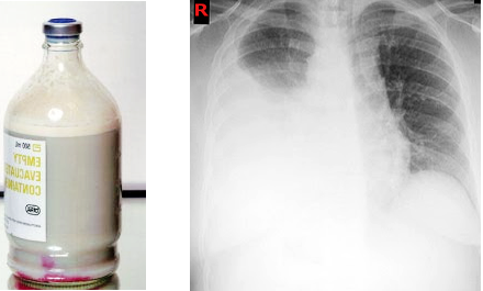
Chest X-Ray Showing Chylothorax With Homogeneous Density.
Contributed by S Bhimji, MD.
References
Schild HH, Pieper C. [Chylothorax: Current Therapeutic Options]. Zentralblatt fur Chirurgie. 2019 Sep:144(S 01):S24-S30. doi: 10.1055/a-0831-2649. Epub 2019 Feb 22 [PubMed PMID: 30795028]
Zhang C, Xi MY, Zeng J, Li Y, Yu C. Prognostic Impact of Postoperative Complications on Overall Survival in 287 Patients With Oral Cancer: A Retrospective Single-Institution Study. Journal of oral and maxillofacial surgery : official journal of the American Association of Oral and Maxillofacial Surgeons. 2019 Jul:77(7):1471-1479. doi: 10.1016/j.joms.2019.01.020. Epub 2019 Jan 26 [PubMed PMID: 30790530]
Level 2 (mid-level) evidenceIio K, Soneda K, Shimotakahara A, Hataya H, Kono T. Effective method of evaluating infantile chylothorax. Pediatrics international : official journal of the Japan Pediatric Society. 2019 Feb:61(2):203-205. doi: 10.1111/ped.13762. Epub 2019 Feb 15 [PubMed PMID: 30767369]
Hayashi K, Hanaoka J, Ohshio Y, Igarashi T. Chylothorax secondary to a pleuroperitoneal communication and chylous ascites after pancreatic resection. Journal of surgical case reports. 2019 Jan:2019(1):rjy364. doi: 10.1093/jscr/rjy364. Epub 2019 Jan 24 [PubMed PMID: 30697412]
Level 3 (low-level) evidenceAgrawal A, Chaddha U, Kaul V, Desai A, Gillaspie E, Maldonado F. Multidisciplinary Management of Chylothorax. Chest. 2022 Dec:162(6):1402-1412. doi: 10.1016/j.chest.2022.06.012. Epub 2022 Jun 20 [PubMed PMID: 35738344]
Cherian S, Umerah OM, Tufail M, Panchal RK. Chylothorax in a patient with HIV-related Kaposi's sarcoma. BMJ case reports. 2019 Jan 22:12(1):. doi: 10.1136/bcr-2018-227641. Epub 2019 Jan 22 [PubMed PMID: 30674495]
Level 3 (low-level) evidenceYamagata Y, Saito K, Hirano K, Oya M. Laparoscopic Transhiatal Thoracic Duct Ligation for Chylothorax after Esophagectomy. The Thoracic and cardiovascular surgeon. 2019 Oct:67(7):606-609. doi: 10.1055/s-0038-1677507. Epub 2019 Jan 22 [PubMed PMID: 30669171]
Gurevich A, Hur S, Singhal S, DiBardino D, Haas AR, Hansen-Flaschen JH, Nadolski G, Itkin M. Nontraumatic Chylothorax and Chylopericardium: Diagnosis and Treatment Using an Algorithmic Approach Based on Novel Lymphatic Imaging. Annals of the American Thoracic Society. 2022 May:19(5):756-762. doi: 10.1513/AnnalsATS.202103-262OC. Epub [PubMed PMID: 34797746]
Dar PMUD, Gamanagatti S, Priyadarshini P, Kumar S. Traumatic chylothorax: a dilemma to surgeons and interventionists. BMJ case reports. 2021 May 21:14(5):. doi: 10.1136/bcr-2020-238961. Epub 2021 May 21 [PubMed PMID: 34020985]
Level 3 (low-level) evidenceMartínez Camacho RJ, Martínez Camacho LI, Martínez Camacho D, Martínez Camacho A. Idiopathic Chylothorax During Pregnancy: A Case Report. Cureus. 2023 Oct:15(10):e47841. doi: 10.7759/cureus.47841. Epub 2023 Oct 27 [PubMed PMID: 37899891]
Level 3 (low-level) evidenceUr Rehman K, Sivakumar P. Non-traumatic chylothorax: diagnostic and therapeutic strategies. Breathe (Sheffield, England). 2022 Jun:18(2):210163. doi: 10.1183/20734735.0163-2021. Epub 2022 Aug 9 [PubMed PMID: 36337134]
Bhatnagar M, Fisher A, Ramsaroop S, Carter A, Pippard B. Chylothorax: pathophysiology, diagnosis, and management-a comprehensive review. Journal of thoracic disease. 2024 Feb 29:16(2):1645-1661. doi: 10.21037/jtd-23-1636. Epub 2024 Feb 22 [PubMed PMID: 38505027]
Papoulidis P, Vidanapathirana P, Dunning J. Chylothorax, new insights in treatment. Journal of thoracic disease. 2018 Nov:10(Suppl 33):S3976-S3977. doi: 10.21037/jtd.2018.09.94. Epub [PubMed PMID: 30631531]
Pospiskova J, Smolej L, Belada D, Simkovic M, Motyckova M, Sykorova A, Stepankova P, Zak P. Experiences in the treatment of refractory chylothorax associated with lymphoproliferative disorders. Orphanet journal of rare diseases. 2019 Jan 9:14(1):9. doi: 10.1186/s13023-018-0991-3. Epub 2019 Jan 9 [PubMed PMID: 30626415]
Yang RF, Liu TT, Wang P, Zhang RQ, Li C, Han B, Gao XX, Zhang L, Jiang ZM. Ligation of thoracic duct during thoracoscopic esophagectomy can lead to decrease of T lymphocyte. Journal of cancer research and therapeutics. 2018:14(7):1535-1539. doi: 10.4103/jcrt.JCRT_596_17. Epub [PubMed PMID: 30589035]
Furukawa M, Hara A, Miyazaki R, Yokoyama S, Hayashi M, Tao H, Okabe K. [Assessment of Fat-free Diet for Postoperative Chylothorax]. Kyobu geka. The Japanese journal of thoracic surgery. 2018 Dec:71(13):1063-1065 [PubMed PMID: 30587742]
Santos LLD, Santos CLD, Hu NKT, Datrino LN, Tavares G, Tristão LS, Orlandini MF, Serafim MCA, Tustumi F. Outcomes of Chylothorax Nonoperative Management After Cardiothoracic Surgery: A Systematic Review and Meta-Analysis. Brazilian journal of cardiovascular surgery. 2023 Oct 6:38(6):e20220326. doi: 10.21470/1678-9741-2022-0326. Epub 2023 Oct 6 [PubMed PMID: 37801640]
Level 1 (high-level) evidenceCosta KM, Saxena AK. Surgical chylothorax in neonates: management and outcomes. World journal of pediatrics : WJP. 2018 Apr:14(2):110-115. doi: 10.1007/s12519-018-0134-x. Epub 2018 Mar 5 [PubMed PMID: 29508361]
Power R, Smyth P, Donlon NE, Nugent T, Donohoe CL, Reynolds JV. Management of chyle leaks following esophageal resection: a systematic review. Diseases of the esophagus : official journal of the International Society for Diseases of the Esophagus. 2021 Nov 11:34(11):. doi: 10.1093/dote/doab012. Epub [PubMed PMID: 33723611]
Level 1 (high-level) evidence