Introduction
Eczema herpeticum is a disseminated cutaneous infection with herpes simplex virus (HSV) that develops in a patient with atopic dermatitis.[1] This condition typically presents as a sudden onset eruption of monomorphic vesicles and "punched-out" erosions with hemorrhagic crusts over eczematous areas. Patients may have systemic symptoms like fever, lymphadenopathy, or malaise.[2] Presentation ranges from mild and self-limiting in healthy adults to life-threatening in children, infants, and immunocompromised patients. Early treatment with antiviral therapy can shorten the duration of mild disease and prevent morbidity and mortality in severe cases.[3]
Etiology
Register For Free And Read The Full Article
 Search engine and full access to all medical articles
- 10 free questions in your specialty
- Free CME/CE Activities
- Free daily question in your email
- Save favorite articles to your dashboard
- Emails offering discounts
Learn more about a Subscription to StatPearls Point-of-Care
Etiology
Eczema herpeticum is due to cutaneous superinfection with herpes simplex virus (HSV), usually HSV-1, in patients with atopic dermatitis. Cases due to reactivation of HSV are more common than primary infection.[4] Individuals with this condition are prone to recurrent bacterial and viral skin infections due to impaired epidermal barrier function and immune dysregulation.[5] Disseminated cutaneous HSV infection can also occur in patients with other forms of dermatitis that impair the skin barrier, which is known as Kaposi varicelliform eruption (KVE). KVE has been reported in patients with Darier disease, Hailey–Hailey disease, immunobullous diseases, burns, irritant contact dermatitis, mycosis fungoides, Sézary syndrome, ichthyoses, and pityriasis rubra pilaris.[6][7]
Risk factors associated with developing eczema herpeticum are more severe atopic skin disease, decreased epidermal expression of filaggrin, and decreased production of cathelicidin and other antimicrobial peptides.[8] Affected patients exhibit biomarkers associated with T-helper type 2 (Th2) cell responses, such as reduced interferon levels, elevated eosinophil count, and increased serum immunoglobulin E levels.[5] The Th2 shift of the immune system in patients with eczema herpeticum is associated with decreased antimicrobial peptides in the epidermis and reduced defense against cutaneous HSV infection.
Eczema herpeticum patients are more likely to have food and environmental allergies, asthma, early onset of atopic dermatitis before age 5, and a history of Staphylococcus aureus and molluscum contagiosum infections.[5] The HLA-B7 allele is associated with an increased risk of eczema herpeticum.[9] Increased interleukin (IL)-10 and local IL-25 expression are found in eczema herpeticum patients and may play a role in developing eczema herpeticum.[10]
Epidemiology
Eczema herpeticum occurs in <3% of patients with atopic dermatitis, affecting infants and children more than adults.[5] Atopic dermatitis is the most common chronic inflammatory skin disease in the world, affecting 10% to 20% of children in developed countries and 7% to 10% of adults in the United States (US).[11][12] Given the prevalence of HSV exposure, the comparative rarity of eczema herpeticum in those with atopic dermatitis suggests many host factors play an important role in the development of eczema herpeticum.
Results from a study of pediatric patients with eczema herpeticum in Canada found that predictors of hospitalization included male sex, age younger than 1 year, fever, and systemic symptoms at presentation.[13] Results from a study conducted in the US found that the average age of hospitalized pediatric patients with eczema herpeticum was 3.26 +/- 0.1 years, and 41.8% of patients were girls. Results from this study also reported higher prevalence, length of hospital stay, and cost of care in Asian pediatric patients.[13] The epidemiology of eczema herpeticum in nonhospitalized and adult patients is not well-defined.
Pathophysiology
The pathophysiology of eczema herpeticum involves a compromised epidermal barrier and dysfunctional immune responses inherent to atopic dermatitis, which predispose the skin to HSV entry and proliferation.[14] In atopic dermatitis, filaggrin deficiency and cytokine-mediated inflammation weaken the skin's structural integrity, allowing HSV to penetrate more easily. The impaired skin barrier, coupled with dysregulated T-cell responses and decreased production of antimicrobial peptides, diminishes the host's ability to control viral replication. This results in widespread vesiculopustular eruptions, often accompanied by systemic symptoms such as fever and malaise.[15]
Histopathology
Histological examination of eczema herpeticum reveals multinucleated giant cells with intranuclear inclusion bodies, known as Cowdry type A inclusions, within the epidermis. These inclusions are indicative of an HSV cytopathic effect. The epidermis shows extensive ballooning degeneration of keratinocytes and intraepidermal vesicle formation. There is often spongiosis and acantholysis, leading to the formation of intraepidermal and subepidermal blisters. The dermis typically exhibits a perivascular lymphocytic infiltrate, sometimes with the presence of eosinophils due to the underlying atopic dermatitis. Additionally, there may be necrosis of keratinocytes and evidence of viral replication within the lesions. These histopathological features are crucial for diagnosing and differentiating eczema herpeticum from other similar vesiculobullous disorders.[10][16]
History and Physical
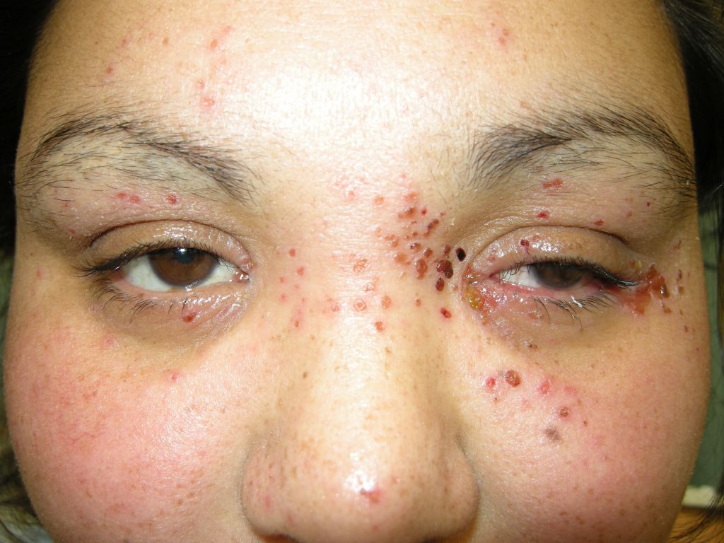
Eczema herpeticum presents as a sudden onset eruption of monomorphic, dome-shaped, grouped, 2 mm to 3 mm vesicles on an erythematous base. Lesions are superimposed on areas of preexisting atopic dermatitis, most commonly on the face, neck, and upper trunk (see Image. Eczema Herpeticum). The lesions are pruritic and painful and may spread to involve normal skin over 7 to 10 days.[17]
Within 2 weeks, the vesicles rupture and form characteristic "punched-out" erosions with the hemorrhagic crust. Small erosions may coalesce to form a larger erosion or ulceration with a scalloped border. Numerous vesicles may appear in successive crops, and multiple morphologies may be present simultaneously. Lesions that are secondarily impetiginized may have an overlying honey-colored crust.
In patients with severe or poorly controlled atopic dermatitis, the characteristic morphology may be difficult to recognize and can be misdiagnosed as an exacerbation of eczematous dermatitis.[18] Patients may have systemic symptoms like fever, malaise, and lymphadenopathy.[3] Eczema herpeticum lesions generally heal without scarring within 6 weeks.[19]
Evaluation
The diagnosis of eczema herpeticum can be made clinically if characteristic morphology is present. Viral polymerase chain reaction (PCR) can be performed on vesicle fluid to confirm the diagnosis and determine the type of herpesvirus with high sensitivity and specificity. If PCR is not available, a Tzank smear, direct fluorescent antibody (DFA) testing, and viral cultures can confirm HSV infection. Bacterial culture should be done if there is a concern for impetiginization. If the clinical presentation is atypical, a skin biopsy may be indicated. Laboratory tests may reveal lymphopenia and an increased erythrocyte sedimentation rate.[17]
Treatment / Management
Patients with eczema herpeticum should be treated promptly with systemic acyclovir or valacyclovir to minimize the risk of complications and prevent progression to severe disease.
- Mild cases can be treated with oral acyclovir or valacyclovir for 7 to 21 days or until all lesions are crusted over.[20] The recommended dosage for oral acyclovir is 30 mg/kg/d to 60 mg/kg/d, divided into 3 doses per day in children and 400 mg 3 times per day in adults.[19][21] The dosage for oral valacyclovir is 20 mg/kg/d in children and 500 mg 3 times per day in adults.[4]
- Severe cases or those who are immunocompromised should be hospitalized for intravenous acyclovir (5 mg/kg to 10 mg/kg every 8 hours). Once clinical improvement occurs and the lesions start to crust over, patients can be transitioned to oral acyclovir.[18] Supportive care with gentle emollients and cool compresses can provide symptomatic relief.
- Those who are critically ill may need intravenous fluids, electrolyte repletion, wound care, pain control, and nutritional support. Patients should be counseled about the risk of autoinoculation, and frequent handwashing should be encouraged. Eczema herpeticum lesions are considered infectious until crusted over.
- Contact precautions should be initiated for those who are hospitalized, including patient isolation and the use of face masks and gowns for healthcare professionals.[21] Patients should be monitored for the development of secondary bacterial infections and treated with systemic antibiotics according to culture and susceptibility results.[21] (B2)
Differential Diagnosis
The differential diagnosis for eczema herpeticum includes the following:
- Impetigo
- Hand-foot-and-mouth disease
- Eczema coxsackium
- Primary varicella infection
- Disseminated herpes zoster
- Disseminated molluscum contagiosum
- Acute generalized exanthematous pustulosis
- Dermatitis herpetiformis
- Cellulitis
- Erysipelas
Misdiagnosis of eczema herpeticum can lead to delayed initiation of antiviral treatment and subsequent complications. Diagnostic clues that favor eczema herpeticum are painful lesions, monomorphic size of the lesions, and characteristic "punched-out" erosions in areas of preexisting atopic dermatitis. Unlike herpes zoster, eczema herpeticum does not respect dermatomal boundaries.
Prognosis
Eczema herpeticum is a potentially life-threatening disease with mortality risk due to complications of systemic viremia, bacteremia, and fungal infection leading to multiorgan failure.[22] Before using acyclovir, mortality rates in patients with eczema herpeticum were reportedly 10% to 50%.[23] Since the widespread implementation of systemic antiviral treatment, mortality rates have decreased significantly.
Results from a 2011 study of 1331 pediatric eczema herpeticum patients hospitalized in the US found no deaths and concluded that the mortality rate of hospitalized patients is low. The median length of hospital stay was 3 days, with 9.2% of patients requiring hospitalization longer than 1 week and 3.8% requiring intensive care unit admission.[24]
Results from a study conducted in 2018 showed that 4655 hospitalized children with eczema herpeticum had a mortality rate of 0.1%, and 98.1% of patients were classified as having a minor mortality risk. Only 4.5% of patients were classified as having a major loss of function, while 33.1% had moderate, and the majority had a minor loss of function.[13]
Complications
Potential complications of eczema herpeticum include cutaneous superinfection with Staphylococcus aureus, Streptococcus pyogenes, and molluscum contagiosum virus.[25][26] Results from a study by Aronson et al found S aureus infection in 30.3% of patients who were pediatric and hospitalized with pediatric eczema, with 9.2% due to methicillin-resistant S aureus and 3.9% with bacteremia. Eighty-six percent of patients were treated with oral or intravenous antibiotics.[24] Additional complications include meningoencephalitis and herpetic keratoconjunctivitis, which can result in scarring and blindness.[4] Disseminated systemic infections of HSV leading to bone marrow suppression, disseminated intravascular coagulation, and death have been reported.[18]
Consultations
A dermatology consult may be beneficial to confirm the diagnosis of eczema herpeticum. An ophthalmologic evaluation is warranted in cases involving the face and eyelids.
Deterrence and Patient Education
Effective deterrence and patient education are crucial in managing eczema herpeticum. Preventing outbreaks involves educating patients and caregivers about proper skin care routines, recognizing early signs of infection, and understanding the importance of prompt medical attention. Patients with atopic dermatitis should be informed about the risk factors for eczema herpeticum, such as severe or poorly controlled dermatitis, and the need to avoid potential triggers, including certain allergens and skin irritants. Healthcare professionals should emphasize the significance of maintaining a robust skincare regimen, using prescribed medications, and attending regular follow-up appointments.
Patients with eczema herpeticum should be counseled that they are infectious until all lesions have crusted over and thus should avoid close contact with others until then. Affected individuals must be encouraged to avoid scratching and should wash their hands frequently due to the risk of autoinoculation. Patients diagnosed with mild eczema herpeticum and treated as outpatients should be cautioned to seek emergency care if they develop systemic symptoms or worsening rash, as they may require hospitalization, intravenous acyclovir treatment, or antibiotic coverage. Immediate treatment can significantly reduce the incidence and severity of eczema herpeticum, ultimately improving patient outcomes and quality of life.
Pearls and Other Issues
Understanding key clinical insights is essential for effectively managing eczema herpeticum. Some clinical pearls that help guide diagnosis, treatment, and patient education include the following:
- Early recognition: Identify eczema herpeticum by its characteristic sudden-onset vesicular rash and "punched-out" erosions, often overlying areas of atopic dermatitis.
- Prompt treatment: Initiate systemic antiviral therapy immediately upon suspicion to reduce morbidity and prevent complications.
- Risk factors: Patients with severe atopic dermatitis, frequent allergies, asthma, early-onset dermatitis, and a history of skin infections are at higher risk.
- Differential diagnosis: Differentiate eczema herpeticum from bacterial infections, impetigo, and other viral exanthems.
- Systemic symptoms: Look for systemic signs such as fever, lymphadenopathy, and malaise, which may accompany the skin findings.
- Secondary infections: Monitor for and treat any secondary bacterial infections promptly.
- Patient education: Educate patients and caregivers on the importance of early symptom recognition and seeking immediate medical care.
- Preventive measures: Encourage strict adherence to prescribed skincare regimens to maintain the skin barrier function and reduce the risk of HSV infection.
- Care coordination: Ensure seamless communication and coordination among interprofessional healthcare team members to effectively manage and follow up on affected patients.
- Follow-up care: Schedule regular follow-up appointments to monitor patient progress and adjust treatment plans as necessary.
Enhancing Healthcare Team Outcomes
Managing eczema herpeticum requires an interprofessional team of physicians, advanced care practitioners, nurses, pharmacists, and other healthcare professionals. Each team member is responsible for specific clinical roles. Physicians and advanced care practitioners must develop skills to promptly recognize the clinical signs of eczema herpeticum, such as uniform vesicles and "punched-out" erosions. Advanced knowledge in prescribing and managing systemic antiviral therapies is crucial for physicians, nurse practitioners, and pharmacists. Nurses should be adept in wound care management, patient monitoring, and educating patients on symptom recognition and care strategies.
The interprofessional team is critical in implementing protocols for the early detection and treatment of eczema herpeticum to prevent adverse effects and complications. Developing and executing integrated care plans that involve multiple healthcare professionals will address all aspects of the patient's condition. Collaborating and establishing effective communication channels is important to ensure comprehensive care while reducing morbidity and mortality associated with eczema herpeticum.
Affected patients and their families should be educated about their condition, treatment options, and prevention strategies. Coordinating follow-up care to monitor the patient's progress and address any emerging issues is essential. By fostering a collaborative, patient-centered approach, interprofessional healthcare teams can improve the quality of care for patients with eczema herpeticum, ultimately leading to better health outcomes and enhanced team performance.
Media
(Click Image to Enlarge)
References
Micali G, Lacarrubba F. Eczema Herpeticum. The New England journal of medicine. 2017 Aug 17:377(7):e9. doi: 10.1056/NEJMicm1701668. Epub [PubMed PMID: 28813215]
Seegräber M, Worm M, Werfel T, Svensson A, Novak N, Simon D, Darsow U, Augustin M, Wollenberg A. Recurrent eczema herpeticum - a retrospective European multicenter study evaluating the clinical characteristics of eczema herpeticum cases in atopic dermatitis patients. Journal of the European Academy of Dermatology and Venereology : JEADV. 2020 May:34(5):1074-1079. doi: 10.1111/jdv.16090. Epub 2020 Jan 30 [PubMed PMID: 31733162]
Level 2 (mid-level) evidenceVera-Kellet C, Hasbún C. Eczema herpeticum: A medical emergency in patients with atopic dermatitis. IDCases. 2020:19():e00663. doi: 10.1016/j.idcr.2019.e00663. Epub 2019 Nov 6 [PubMed PMID: 32226756]
Level 3 (low-level) evidenceWollenberg A, Wetzel S, Burgdorf WH, Haas J. Viral infections in atopic dermatitis: pathogenic aspects and clinical management. The Journal of allergy and clinical immunology. 2003 Oct:112(4):667-74 [PubMed PMID: 14564342]
Leung DY. Why is eczema herpeticum unexpectedly rare? Antiviral research. 2013 May:98(2):153-7. doi: 10.1016/j.antiviral.2013.02.010. Epub 2013 Feb 22 [PubMed PMID: 23439082]
Lehman JS, el-Azhary RA. Kaposi varicelliform eruption in patients with autoimmune bullous dermatoses. International journal of dermatology. 2016 Mar:55(3):e136-40. doi: 10.1111/ijd.13091. Epub 2015 Oct 24 [PubMed PMID: 26500144]
Walker K, Martini A, Philips H, Sharp L, Thomas K, Tarbox M. Darier disease with disseminated herpes simplex virus type 2 infection. Dermatology online journal. 2019 Apr 15:25(4):. pii: 13030/qt4qg8s971. Epub 2019 Apr 15 [PubMed PMID: 31046908]
Hata TR, Kotol P, Boguniewicz M, Taylor P, Paik A, Jackson M, Nguyen M, Kabigting F, Miller J, Gerber M, Zaccaro D, Armstrong B, Dorschner R, Leung DY, Gallo RL. History of eczema herpeticum is associated with the inability to induce human β-defensin (HBD)-2, HBD-3 and cathelicidin in the skin of patients with atopic dermatitis. The British journal of dermatology. 2010 Sep:163(3):659-61. doi: 10.1111/j.1365-2133.2010.09892.x. Epub 2010 Jun 9 [PubMed PMID: 20545685]
Level 3 (low-level) evidenceMathias RA, Weinberg A, Boguniewicz M, Zaccaro DJ, Armstrong B, Schneider LC, Hata TR, Hanifin JM, Beck LA, Barnes KC, Leung DY. Atopic dermatitis complicated by eczema herpeticum is associated with HLA B7 and reduced interferon-γ-producing CD8+ T cells. The British journal of dermatology. 2013 Sep:169(3):700-3. doi: 10.1111/bjd.12382. Epub [PubMed PMID: 23600999]
Level 2 (mid-level) evidenceDamour A, Garcia M, Seneschal J, Lévêque N, Bodet C. Eczema Herpeticum: Clinical and Pathophysiological Aspects. Clinical reviews in allergy & immunology. 2020 Aug:59(1):1-18. doi: 10.1007/s12016-019-08768-3. Epub [PubMed PMID: 31836943]
Lyons JJ, Milner JD, Stone KD. Atopic dermatitis in children: clinical features, pathophysiology, and treatment. Immunology and allergy clinics of North America. 2015 Feb:35(1):161-83. doi: 10.1016/j.iac.2014.09.008. Epub 2014 Nov 21 [PubMed PMID: 25459583]
Silverberg JI, Hanifin JM. Adult eczema prevalence and associations with asthma and other health and demographic factors: a US population-based study. The Journal of allergy and clinical immunology. 2013 Nov:132(5):1132-8. doi: 10.1016/j.jaci.2013.08.031. Epub 2013 Oct 4 [PubMed PMID: 24094544]
Hsu DY, Shinkai K, Silverberg JI. Epidemiology of Eczema Herpeticum in Hospitalized U.S. Children: Analysis of a Nationwide Cohort. The Journal of investigative dermatology. 2018 Feb:138(2):265-272. doi: 10.1016/j.jid.2017.08.039. Epub 2017 Sep 18 [PubMed PMID: 28927889]
Traidl S, Roesner L, Zeitvogel J, Werfel T. Eczema herpeticum in atopic dermatitis. Allergy. 2021 Oct:76(10):3017-3027. doi: 10.1111/all.14853. Epub 2021 May 3 [PubMed PMID: 33844308]
Branco EA, Ribeiro L, Ribeiro M, Martins A, Policarpo S, Enes J, Silva JML, Santos A, Sarmento A, Ceia F. Eczema herpeticum. IDCases. 2021:26():e01299. doi: 10.1016/j.idcr.2021.e01299. Epub 2021 Oct 1 [PubMed PMID: 34660201]
Level 3 (low-level) evidenceShah VV, Fischer R, Squires S, Tonkovic-Capin V. Eczema herpeticum in a patient With Hailey-Hailey disease confounded by coexistent psoriasis. Cutis. 2020 Jun:105(6):E38-E41 [PubMed PMID: 32717004]
Wollenberg A, Zoch C, Wetzel S, Plewig G, Przybilla B. Predisposing factors and clinical features of eczema herpeticum: a retrospective analysis of 100 cases. Journal of the American Academy of Dermatology. 2003 Aug:49(2):198-205 [PubMed PMID: 12894065]
Level 2 (mid-level) evidenceZhuang K, Wu Q, Ran X, Ran Y, Ding L, Xu X, Lei S, Lama J. Oral treatment with valacyclovir for HSV-2-associated eczema herpeticum in a 9-month-old infant: A case report. Medicine. 2016 Jul:95(29):e4284. doi: 10.1097/MD.0000000000004284. Epub [PubMed PMID: 27442669]
Level 3 (low-level) evidenceLuca NJ, Lara-Corrales I, Pope E. Eczema herpeticum in children: clinical features and factors predictive of hospitalization. The Journal of pediatrics. 2012 Oct:161(4):671-5. doi: 10.1016/j.jpeds.2012.03.057. Epub 2012 May 9 [PubMed PMID: 22575249]
Level 2 (mid-level) evidenceAtherton DJ, Harper JI. Management of eczema herpeticum. Journal of the American Academy of Dermatology. 1988 Apr:18(4 Pt 1):757-8 [PubMed PMID: 3372774]
Level 3 (low-level) evidenceSohail M, Khan FA, Shami HB, Bashir MM. Management of eczema herpeticum in a Burn Unit. JPMA. The Journal of the Pakistan Medical Association. 2016 Nov:66(11):1357-1361 [PubMed PMID: 27812048]
Wollenberg A. Eczema herpeticum. Chemical immunology and allergy. 2012:96():89-95. doi: 10.1159/000331892. Epub 2012 Mar 13 [PubMed PMID: 22433376]
Wheeler CE Jr, Abele DC. Eczema herpeticum, primary and recurrent. Archives of dermatology. 1966 Feb:93(2):162-73 [PubMed PMID: 4159394]
Level 3 (low-level) evidenceAronson PL, Yan AC, Mittal MK, Mohamad Z, Shah SS. Delayed acyclovir and outcomes of children hospitalized with eczema herpeticum. Pediatrics. 2011 Dec:128(6):1161-7. doi: 10.1542/peds.2011-0948. Epub 2011 Nov 14 [PubMed PMID: 22084327]
Level 2 (mid-level) evidenceBeck LA, Boguniewicz M, Hata T, Schneider LC, Hanifin J, Gallo R, Paller AS, Lieff S, Reese J, Zaccaro D, Milgrom H, Barnes KC, Leung DY. Phenotype of atopic dermatitis subjects with a history of eczema herpeticum. The Journal of allergy and clinical immunology. 2009 Aug:124(2):260-9, 269.e1-7. doi: 10.1016/j.jaci.2009.05.020. Epub 2009 Jun 27 [PubMed PMID: 19541356]
Narla S, Silverberg JI. Association between atopic dermatitis and serious cutaneous, multiorgan and systemic infections in US adults. Annals of allergy, asthma & immunology : official publication of the American College of Allergy, Asthma, & Immunology. 2018 Jan:120(1):66-72.e11. doi: 10.1016/j.anai.2017.10.019. Epub [PubMed PMID: 29273131]