Definition/Introduction
The Lasegue sign or straight leg raise (SLR) test is a clinical test to assess nerve root irritation in the lumbosacral area.[1] This test is an integral part of the neurological examination of the patients presenting with low back pain with or without radicular symptoms. The other less commonly used name is the Lazarevic sign (see Image. Lasegue Sign).
Historical Evolution
Although Ernest-Charles Lasegue (1816-1883) was not the first to publish the description of this maneuver, he is considered the first physician who verbally described this sign and emphasized its importance in patients with sciatica.[2][3] Lazarevic (1851-1891) was the first physician to publish this sign in 1880 with a sound pathophysiological explanation.[3] Later, Frost, a student of Lasegue, also described the sign in his doctoral thesis titled Contribution a` l'e´tude Clinique de la Sciatique in 1881. Lasegue and Frost proposed that the sharp pain elicited by the test was due to compression of the sciatic nerve by muscular contraction.[2] In contrast, Lazarevic suggested sciatic nerve stretching as the cause of pain during the SLR test. The cadaveric experiment performed by Lucien de Beurmann in 1884 supported Lazarevic's explanation.[2][4] Later, several modifications were introduced to the SLR test to reproduce pain in irritated nerve roots. These modifications were intended to improve the accuracy of the test.[5][6] Of note, although there is no general agreement on interpreting the results of the SLR test and its variants, performing a combination of tests can enhance their accuracy.[5][7]
Anatomy Related to the Straight Leg Raise Test
In the lumbar region, the nerve roots cross the intervertebral disc above the neural foramina through which they exit.[6] The neural foramen is bounded by the pedicle superiorly, ligamentum flavum posteriorly, and the vertebral body with disc anteriorly. Within the neural foramen, the nerve roots are surrounded by loose areolar tissue and are lightly tethered to adjacent solid structures. This arrangement allows the nerve roots some room for movement while the limbs move. Recent studies have highlighted that this anatomical arrangement is crucial for understanding nerve root mobility during various physical examinations, including the SLR test. Research has shown that the normal average excursion of lumbosacral nerve roots is about 4 to 6 mm, which decreases with age, affecting test results. This dynamic is particularly relevant when evaluating patients with lumbar disc herniation, where reduced nerve root movement is observed. The implications of this for diagnosing and managing radicular pain have been discussed in recent literature, emphasizing the need for accurate assessment techniques. In patients with disc prolapse, there is a loss of nerve root movement, mainly due to adhesion secondary to the local inflammation and partly due to mechanical compression.[8][9][10] This restriction limits hip flexion with an extended knee compared to normal individuals, such as lumbar disc prolapse. In patients with disc prolapse, there is a loss of nerve root movement, mainly due to adhesion secondary to the local inflammation and partly due to mechanical compression. Both mechanisms work together to reduce the SLR angle. During the SLR test, the tension occurs initially, and then the movement appears distally, followed by proximally, along the course of the sciatic nerve and nerve roots as the hip is flexed.
Causes of Pain While Performing the Straight Leg Raise Test
- Stretching of the sciatic nerve
- Displacement of the medulla and conus medullaris
- Nerve compression leads to sensitization at the dorsal root ganglion and posterior horn, lowering the pain threshold [4]
Causes of Positive Straight Leg Raise Test
- Nerve root irritation: Intervertebral disc prolapse, particularly at the L4-L5 or L5-S1 levels, is the most common cause.
- Intraspinal tumor
- Spinal stenosis
- Spondylolisthesis
- Inflammatory radiculopathy
Examination Techniques
Forst described the original examination method: The patient is supine, and the affected lower limb is raised with the knee extended. This maneuver should reproduce pain. The examiner then repeats the maneuver with the leg flexed at the knee and the thigh flexed on the pelvis. This adjustment should not evoke pain.[2][6]
The following technique is currently employed in clinical practice: The patient should be informed about the test steps, what to expect during the exam, and how to describe the pain distribution. The patient should be positioned supine, with the head slightly extended. During the examination, the hips and legs should stay neutral. No hip abduction, adduction, internal or external rotation of the leg is allowed is allowed. The affected leg is then passively and slowly raised by the ankle with the knee fully extended. Upon eliciting pain, the examiner stops further leg elevation and records the range of motion and the area of pain distribution.[6][12]
Recent recommendations emphasize the importance of clear communication with the patient to avoid discomfort and improve test accuracy. Proper positioning and technique are crucial to avoid false positives and ensure reliable results. In addition, maintaining patient comfort can help minimize muscle tension, which might otherwise influence the test outcomes.
Noteworthily, ankle dorsiflexion during the SLR test may exaggerate the pain, although it is not part of the Lasegue sign.[13]
Criteria for a True Positive Straight Leg Raise Test
- Radicular leg pain should occur, radiating below the knee.
- Pain occurs when the leg is between 30° and 60° or 70° from horizontal.[5][6]
Findings That Do Not Qualify as a Positive Straight Leg Raise Test
- Pain occurs in the lower back alone.
- Pain occurs in the posterior thigh alone.
- Pain occurs at an angle less than 30° and may indicate non-organicity or hip joint pathology.
- Pain occurs at an angle of more than 70° from the horizontal, likely due to tight hamstrings or gluteal muscles.
- Pain occurs in a healthy person at an angle of 80° to 90°.
Issues of Concern
Register For Free And Read The Full Article
 Search engine and full access to all medical articles
- 10 free questions in your specialty
- Free CME/CE Activities
- Free daily question in your email
- Save favorite articles to your dashboard
- Emails offering discounts
Learn more about a Subscription to StatPearls Point-of-Care
Issues of Concern
Modifications and Variants of the Straight Leg Raise Test
The accuracy of the SLR test can be improved if it is interpreted along with other clinical tests, including:
- Crossed SLR test: Also known as the well-leg raising test or Fajersztajn sign. When the contralateral leg is lifted, the patient experiences pain on the affected side. This test is more specific than the ipsilateral SLR test and often indicates severe compression or centrally located disc prolapse. Fajersztajn believed that this sign was caused by disc prolapse in the axilla of the root.[6][16][15]
- Reverse SLR test: Also known as the femoral stretch or Ely test. Although the patient is prone, the leg is lifted off the table with both hip and knee joints extended. Some authors may allow knee flexion. This maneuver may reproduce radicular pain in the case of upper lumbar radiculopathy, far lateral lumbar disc pathology, or femoral neuropathy. The pain is elicited in the femoral nerve distribution on the side of the lesion.[17]
- Braggard test: Also known as the sciatic stretch test or flip test. When raising the leg, the foot is held in a dorsiflexed position so that the sciatic nerve is stretched more, increasing the pain intensity or making it possible to elicit the sign early.[18][19]
- Reverse flip test: When raising the leg, the foot is held in a plantar-flexed position, reducing the pain. An increase in pain in this position may suggest malingering.
- Bowstring sign: Also known as the popliteal compression test or posterior tibial nerve stretch sign. The examiner flexes the knee and applies pressure on the popliteal fossa, evoking sciatica. Some examiners perform this maneuver after the SLR test by flexing the knee to relieve the buttock pain. A quick snap on the posterior tibial nerve in the popliteal fossa can reproduce the pain.[15]
Other Nerve Root Irritation Tests
The following clinical tests may also be used to test for nerve root irritation, although their use is less frequent:
- Sitting SLR test (Bechterew test): The patient is made to sit at the edge of a table with both hip and knee flexed, then extend the knee joint or elevate the extended knee, reproducing the radicular pain. The patient may be able to extend each leg alone, but extending both causes radicular pain.[20]
- Distracted SLR test: A sitting SLR test is performed. The patient is distracted as if the physician is examining the foot or pulsation, and slowly, the knee is extended. If the patient is experiencing true radiculopathy, the same pain is reproduced.
- Neri sign: Although bending forward, the patient flexes the knee to avoid stretching the nerve.[19]
- Buckling sign: The patient may flex the knee during the SLR test to avoid sciatic nerve tension.[19]
- Sicard sign: Passive dorsiflexion of the ipsilateral great toe just at the angle of the SLR test produces more pain.
- Kraus-Weber test: The patient may be able to do a sit-up with the knees flexed but not extended.
- Minor sign: The patient may rise from a seated position by supporting themselves on the unaffected side, bending forward, and placing 1 hand on the affected side of the back.
- Bonnet phenomenon: The pain may be more severe or elicited sooner if the test is carried out with the thigh and leg in a position of adduction and internal rotation.
Clinical Significance
Interpretation of the Straight Leg Raise Test
- L4 nerve root irritation: Pain radiating down the buttock to the lateral thigh and medial calf.
- L5 nerve root irritation: Pain radiating down the buttock to the posterior thigh and lateral calf.
- S1 nerve root irritation: Pain radiating down the buttock to the posterior thigh, calf, and lateral foot.
Interpretation of a Positive Reverse Straight Leg Raise Test
- L2, L3, or L4 root irritation
- Femoral nerve irritation
Sensitivity and Specificity of the Test
The sensitivity of the SLR for diagnosing conditions such as herniation of the lumbar disc or irritation of the sciatic nerve varies based on the condition and the study. The sensitivity of the ipsilateral SLR is 72% to 97%, and specificity is 11% to 66%. In contrast, the sensitivity of the crossed SLR test is 23% to 42%, which is less than that of the ipsilateral SLR test but more specific (85% to 100%).[21] The SLR is often combined with other tests and imaging to improve diagnostic accuracy in the clinical setting.
Tests to Confirm Non-organicity While Performing the Straight Leg Raise Test
- Pain occurring at an angle less than 30°
- A significant discrepancy between the supine and sitting SLR test
- Touch-me-not or Waddell sign: Widespread and excessive tenderness
- Back pain when pressing down on the top of the head
- Overreaction during testing
- Non-dermatomal and non-myotomal neurologic signs
- Pain during simulated spinal rotation: The patient's hands remain to the sides, and the hips are rotated. This maneuver does not rotate the spine, but the patient may still report pain.
Nursing, Allied Health, and Interprofessional Team Interventions
When performing the SLR test on a female patient, a clinical chaperone is essential. Patients wearing saris or skirts may feel inhibited to raise their legs, which can lead to resistance in the movement and further misinterpretation of the SLR test; such hesitation is reasonably avoidable with the presence of a chaperone. Counseling and reassurance are crucial in cases where the patient is in significant pain. The patient should be informed about the examination steps and reassured that their comfort is a priority. Proper communication throughout the test can help manage their anxiety and prevent unnecessary pain. The examiner must take great care to position the patient without exacerbating their discomfort, adjusting the test to ensure patient safety and well-being. Effective communication between the examining clinician and chaperone is critical to performing the SLR test accurately. This collaboration helps reduce the likelihood of false positives, as the clinical chaperone can assist with positioning and observation, ensuring that the test is conducted smoothly and without unnecessary strain on the patient. Involving the chaperone in the process promotes a patient-centered approach, ensuring the test is performed with empathy and professionalism.
Media
(Click Image to Enlarge)
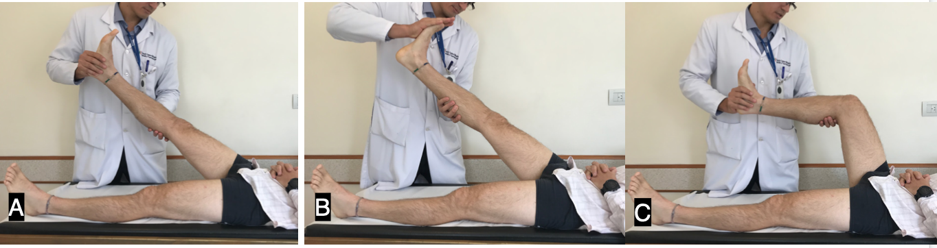
Lasegue Sign. The images show the straight leg raise test (A) and the Braggard test, which increases the sensitivity of the test. (B) The patient typically experiences pain relief when the knee is flexed.
Contributed by G Camino Willhuber, MD
References
Boyd BS, Topp KS, Coppieters MW. Impact of movement sequencing on sciatic and tibial nerve strain and excursion during the straight leg raise test in embalmed cadavers. The Journal of orthopaedic and sports physical therapy. 2013 Jun:43(6):398-403. doi: 10.2519/jospt.2013.4413. Epub 2013 Apr 30 [PubMed PMID: 23633619]
WARTENBERG R. On neurologic terminology, eponyms and the Lasègue sign. Neurology. 1956 Dec:6(12):853-8 [PubMed PMID: 13378588]
Maranhão-Filho P, Vincent M. Lazarević-Lasègue sign. Arquivos de neuro-psiquiatria. 2018 Jun:76(6):421-423. doi: 10.1590/0004-282X20180050. Epub [PubMed PMID: 29972425]
Rade M, Könönen M, Marttila J, Vanninen R, Shacklock M, Kankaanpää M, Airaksinen O. Correlation analysis of demographic and anthropometric factors, hip flexion angle and conus medullaris displacement with unilateral and bilateral straight leg raise. European spine journal : official publication of the European Spine Society, the European Spinal Deformity Society, and the European Section of the Cervical Spine Research Society. 2016 Mar:25(3):724-31. doi: 10.1007/s00586-015-3861-x. Epub 2015 Mar 13 [PubMed PMID: 25763871]
Dyck P. Lumbar nerve root: the enigmatic eponyms. Spine. 1984 Jan-Feb:9(1):3-6 [PubMed PMID: 6372123]
Supik LF, Broom MJ. Sciatic tension signs and lumbar disc herniation. Spine. 1994 May 1:19(9):1066-9 [PubMed PMID: 8029743]