Introduction
Miliaria, also known as eccrine miliaria, is a common skin condition caused by the blockage of eccrine sweat glands and ducts, leading to the backflow of eccrine sweat into the dermis or epidermis.[1] This backflow results in a rash comprising sweat-filled vesicle formation under the skin.
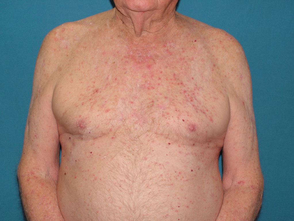
The 3 main types of miliaria—crystallina, rubra, and profunda—are classified by the depth of sweat duct obstruction, which leads to different clinical and histological presentations. Miliaria is often referred to as "heat rash," "prickly heat," or "sweat rash."[2] This condition is most prevalent in warm, humid climates, especially in summer. The rash is typically self-limiting and usually resolves without treatment (see Image. Miliaria, Skin Disease).
Etiology
Register For Free And Read The Full Article
 Search engine and full access to all medical articles
- 10 free questions in your specialty
- Free CME/CE Activities
- Free daily question in your email
- Save favorite articles to your dashboard
- Emails offering discounts
Learn more about a Subscription to StatPearls Point-of-Care
Etiology
Although miliaria affects all age groups, ethnicities, and both genders equally, infants and children are at higher risk due to the immaturity of their eccrine ducts.[3] Sweating is the most common risk factor, making hot or humid conditions and high fevers particularly associated with the development of miliaria.
Other causes of miliaria include:
- Occlusion of the skin: Transdermal drug patches and tight clothing have been associated with miliaria.[4][5]
- Type I pseudohypoaldosteronism: This condition involves mineralocorticoid resistance, leading to sodium loss through eccrine glands, and has been associated with pustular miliaria rubra.[6][7][8]
- Strenuous physical activity: Intense exercise can contribute to the development of miliaria.
- Morvan syndrome: A rare autosomal recessive disorder characterized by hyperhidrosis, among other abnormalities, which predisposes individuals to miliaria.[9][10]
- Medications: Drugs that induce sweating, such as bethanechol, clonidine, and neostigmine, have been associated with miliaria.[11] Additionally, a few cases of isotretinoin-induced miliaria have been reported.[12]
Epidemiology
Miliaria frequently affects neonates and adults with increased sweating, as well as individuals living in hot and humid climates. While miliaria can occur in both genders and across all ethnicities, each type may be more common in specific populations.
- Miliaria crystalline: This condition is also known as sudamina, and it commonly affects neonates, with the highest incidence observed at 2 weeks of age or younger.[13] The rash affects approximately 4.5% to 9% of neonates.[14][15] However, it can also occur in adults who have recently relocated to a warmer climate.[16]
- Miliaria rubra: This is the most common form of miliaria and is frequently observed in neonates between 1 and 3 weeks of age. This condition can also affect up to 30% of adults living in hot and humid conditions.[3]
- Miliaria profunda: This is the rarest form of miliaria, most commonly seen in individuals with recurrent episodes of miliaria rubra or individuals exposed to new warm climates, such as military personnel deployed in tropical climates.[17]
Pathophysiology
The main cause of miliaria is the obstruction of eccrine sweat glands or ducts. This obstruction can result from cutaneous debris or bacteria, such as S epidermidis, which forms biofilms.[18][19] The obstruction leads to sweat leakage into the epidermis or dermis, causing cellular overhydration, swelling, and further occlusion of the ducts. In more severe cases, deeper involvement of the eccrine glands or ducts may lead to their rupture.
The different types of miliaria involve varying depths of cutaneous obstruction. Miliaria crystallina occurs with ductal occlusion in the stratum corneum; miliaria rubra involves ductal occlusion in the epidermis at the subcorneal layers; and miliaria profunda results from ductal occlusion at the dermal-epidermal junction, specifically within the papillary dermis.
Histopathology
The histology of miliaria varies by type, as each is classified by the depth of obstruction in the eccrine duct.
- In miliaria crystallina, histology reveals subcorneal or intracorneal vesicles originating from the intraepidermal portion of the duct, which may also contain neutrophils.
- Miliaria rubra is characterized by epidermal spongiosis with parakeratosis and vesicles within the epidermis that communicate with the eccrine duct. This condition may also be associated with an inflammatory lymphocytic infiltrate surrounding the duct and superficial vasculature.
- Miliaria profunda involves intradermal spongiosis of the eccrine duct and is similar to miliaria rubra. However, miliaria profunda differs from miliaria rubra by involving more extensive rupture of the eccrine ducts and more pronounced lymphocytic inflammation. This also shows periodic acid-Schiff positivity and is diastase-resistant under microscopy.
History and Physical
A comprehensive medical history should be obtained, as it is essential for accurately diagnosing and effectively managing miliaria. Clinicians should begin by inquiring about the onset, duration, and progression of skin symptoms, including the appearance and location of vesicles, papules, or pustules. Notably, it is important to inquire about recent environmental changes, such as relocation to a hotter or more humid climate or increased sweating due to physical activity or illness. The interprofessional healthcare team should also gather information on any recent exposure to irritants or new skincare products, review the patient's medical history for conditions that might affect sweat gland function, and check for any previous episodes of miliaria. Understanding these factors is crucial for distinguishing miliaria from other skin conditions and guiding appropriate treatment.
Clinical Examination of Miliaria
Miliaria is characterized by the presence of vesicles, papules, and pustules on the skin. A thorough examination of the skin, focusing on the characteristics and location of the rash, is essential for an accurate diagnosis.
Miliaria crystalline: This condition appears as 1 to 2 mm superficial vesicles, affecting both adults and neonates, usually younger than 2 weeks old.[20] The pathophysiology involves the most superficial layer of the epidermis, the stratum corneum, resulting in vesicles with a thin superficial layer that resembles water droplets on the skin and easily ruptures.[11][21] As these vesicles are so superficial, an inflammatory response is typically absent. The most commonly affected areas are the upper trunk, neck, and head. The rash usually appears within a few days of exposure to risk factors and resolves within a day after the superficial layer of skin rubs off.[22]
Miliaria rubra: This condition is the most prevalent form of miliaria and is characterized by the obstruction of eccrine ducts in the deeper layers of the skin, which triggers an inflammatory response. This leads to the formation of larger, erythematous papules and vesicles. A key clinical feature that helps differentiate miliaria rubra from folliculitis is the minimal involvement of hair follicles. When pustules are present, miliaria rubra is referred to as miliaria pustulosa, which may indicate a bacterial infection. Due to the inflammatory response, patients may experience pruritic and painful symptoms that can worsen during perspiration, leading to increased irritation. In neonates, typically between 1 and 3 weeks old, the groin, axilla, and neck are the most commonly affected areas. In adults, miliaria rubra is often seen in areas where clothing rubs against the skin, such as the trunk and extremities, while the face is usually spared. Superinfection with staphylococci can occur, and when associated with impetigo or multiple abscesses, the condition is known as periporitis staphylogenes.[23]
Miliaria profunda: Miliaria profunda results in large, firm, flesh-colored papules that are not centered around hair follicles due to their deeper involvement at the dermal-epidermal junction. The eruption can range from extremely pruritic to asymptomatic. This condition is often observed in patients with a history of multiple episodes of miliaria rubra. The rash typically affects the trunk in adults, but it may also involve the arms and legs. The rash usually appears within minutes to hours of sweating and resolves within an hour after sweating ceases.
In both miliaria rubra and miliaria profunda, anhidrosis may occur in the affected areas due to obstructed sweat glands. Consequently, heat exhaustion can result from ineffective thermoregulation in patients with extensive involvement, and this should be considered when a skin rash is accompanied by hyperthermia.
Evaluation
Miliaria is primarily diagnosed clinically, as laboratory tests are often inconclusive and generally not useful. Dermoscopy is a valuable tool, particularly for individuals with darker skin, as it can reveal large white globules with surrounding darker halos (white bullseye).[24] If the diagnosis remains uncertain, a skin punch biopsy may be helpful. High-definition optical coherence tomography can also assist in identifying the obstruction of the eccrine duct.[25]
Treatment / Management
As hot and sweaty conditions are primary risk factors for miliaria, managing the condition involves implementing general measures to reduce sweating and prevent eccrine duct blockage. This includes creating cooler environments, wearing breathable clothing, exfoliating the skin, removing skin-occluding objects such as bandages or patches, and addressing febrile illnesses.
Specific modalities for the treatment of miliaria are unique depending on the type. Miliaria crystallina usually does not require treatment, as it is self-limiting and typically resolves within 24 hours. Miliaria rubra is managed by reducing inflammation; thus, mild-to-mid-potency corticosteroids, such as 0.1% triamcinolone cream, may be applied to the affected areas for 1 to 2 weeks. If miliaria pustulosa develops, topical antibiotics such as clindamycin are used to address the superimposed bacterial infection.
Limited information exists on the treatment of miliaria profunda beyond the general measures previously mentioned. However, studies have indicated that miliaria profunda may improve with a combined regimen of oral isotretinoin (40 mg/d for 2 months) and topical anhydrous lanolin.[26](B3)
Differential Diagnosis
Miliaria clinically resembles and must be differentiated from the following:
- Viral exanthems or viral infections, such as herpes simplex or varicella
- Cutaneous candidiasis or other fungal skin infections
- Folliculitis, whether bacterial or pityrosporum
- Neonatal acne or erythema toxicum neonatorum
- Drug rashes, particularly acute, generalized, exanthematous pustulosis
- Grover disease
- Arthropod bites
- Lymphocytoma cutis or cutaneous T-cell pseudolymphomas
Prognosis
Most cases of miliaria resolve spontaneously once risk factors are reduced and the patient is moved to a cooler, less humid environment. All types of miliaria typically resolve relatively easily, except for miliaria profunda, which can have a variable prognosis. While symptoms often improve with treatment, individuals may be prone to recurrent episodes, especially in hot climates. Chronic cases can potentially cause permanent damage to sweat glands, leading to anhidrosis and an increased risk of heat-related illnesses.
Complications
The most severe complication of miliaria, particularly miliaria profunda, is anhidrosis, which impairs thermoregulation and can lead to heat exhaustion. This can permanently impact a person's ability to work or prevent an active individual from continuing exercise or sports. Opportunistic bacterial superinfections may also occur in miliaria pustulosa and rubra due to changes in the affected epidermal layer caused by this skin condition.
Consultations
Consultations may be necessary for patients with miliaria, especially in cases where the condition is severe, recurrent, or resistant to standard treatments. Dermatologists should be consulted to confirm the diagnosis, particularly when miliaria resembles other dermatoses, or there is uncertainty in differentiating between its types. For patients with underlying health conditions that exacerbate miliaria, such as hyperhidrosis or autoimmune disorders, a multidisciplinary approach involving endocrinologists or immunologists may be warranted. In cases where miliaria is related to environmental or occupational factors, consultations with occupational health specialists can offer guidance on modifying the patient's environment to prevent future outbreaks. Timely consultations ensure a comprehensive treatment plan tailored to the patient's specific needs and circumstances.
Deterrence and Patient Education
Deterrence and patient education are crucial in managing and preventing miliaria, especially for those prone to the condition. Patients should be informed about the importance of avoiding excessive heat and humidity, which can trigger or worsen miliaria. They should be encouraged to wear loose-fitting, breathable clothing and to stay in cool, air-conditioned environments whenever possible. Additionally, affected individuals should be advised on proper skincare, including gentle cleansing and avoiding heavy creams or ointments that can block sweat ducts. Emphasizing the need to stay hydrated and take frequent cool showers to reduce sweat buildup is also important.
Early intervention, such as cooling the skin at the first sign of irritation or rash, can help prevent miliaria from progressing and becoming more severe. The clinician should develop a personalized care plan for the patient and educate them on recognizing the signs and symptoms of heat exhaustion, especially if anhidrosis occurs due to miliaria.
Enhancing Healthcare Team Outcomes
An interprofessional healthcare team, including primary clinicians, advanced care practitioners, nurses, and pharmacists, is critical in improving patient outcomes with miliaria. Clinicians must accurately diagnose miliaria, differentiate it from other skin conditions, and understand its pathophysiology. Effective management requires knowledge of appropriate treatments, such as topical therapies, and preventive strategies, such as environmental modifications. Additionally, skills in patient education are essential to ensure that patients understand how to manage their condition and prevent future outbreaks.
Healthcare professionals should collaborate to develop individualized care plans that address each patient’s needs, including considerations for age, underlying health conditions, and environmental exposures. Preventive strategies, such as advising on climate control and proper skin care, are crucial for minimizing recurrences. If a rash that appears to be miliaria does not resolve promptly, a referral to a dermatologist is recommended to rule out other more severe conditions that may mimic miliaria.
Coordinated care ensures that all treatments are compatible, follow-up appointments are scheduled, and patient education is consistent across the healthcare team. This is particularly crucial for patients with complex or recurrent miliaria, where multiple specialties may be involved. An interprofessional approach enhances patient-centered care, improves outcomes, ensures patient safety, and optimizes team performance in treating affected individuals.
Media
(Click Image to Enlarge)
References
Wenzel FG, Horn TD. Nonneoplastic disorders of the eccrine glands. Journal of the American Academy of Dermatology. 1998 Jan:38(1):1-17; quiz 18-20 [PubMed PMID: 9448199]
El Anzi O, Hassam B. [Widespread miliaria crystallina: about a case]. The Pan African medical journal. 2018:30():69. doi: 10.11604/pamj.2018.30.69.15383. Epub 2018 May 28 [PubMed PMID: 30344853]
Level 3 (low-level) evidenceLYONS RE, LEVINE R, AULD D. Miliaria rubra, a manifestation of staphylococcal disease. Archives of dermatology. 1962 Sep:86():282-6 [PubMed PMID: 14467655]
Ale I, Lachapelle JM, Maibach HI. Skin tolerability associated with transdermal drug delivery systems: an overview. Advances in therapy. 2009 Oct:26(10):920-35. doi: 10.1007/s12325-009-0075-9. Epub 2009 Nov 27 [PubMed PMID: 19967501]
Level 3 (low-level) evidenceCarter R 3rd, Garcia AM, Souhan BE. Patients presenting with miliaria while wearing flame resistant clothing in high ambient temperatures: a case series. Journal of medical case reports. 2011 Sep 22:5():474. doi: 10.1186/1752-1947-5-474. Epub 2011 Sep 22 [PubMed PMID: 21939537]
Level 2 (mid-level) evidenceOnal H, Adal E, Ersen A, Onal Z, Keskindemirci G. Miliaria rubra and thrombocytosis in pseudohypoaldosteronism: case report. Platelets. 2012:23(8):645-7. doi: 10.3109/09537104.2011.641624. Epub 2011 Dec 13 [PubMed PMID: 22150373]
Level 3 (low-level) evidenceUrbatsch A, Paller AS. Pustular miliaria rubra: a specific cutaneous finding of type I pseudohypoaldosteronism. Pediatric dermatology. 2002 Jul-Aug:19(4):317-9 [PubMed PMID: 12220275]
Level 3 (low-level) evidenceAkcakus M, Koklu E, Poyrazoglu H, Kurtoglu S. Newborn with pseudohypoaldosteronism and miliaria rubra. International journal of dermatology. 2006 Dec:45(12):1432-4 [PubMed PMID: 17184247]
Level 3 (low-level) evidenceTabanelli M, Passarini B, Liguori R, Balestri R, Gaspari V, Giacomini F, Patrizi A. Erythematous papules on the parasternal region in a 76-year-old man. Clinical and experimental dermatology. 2008 May:33(3):369-70. doi: 10.1111/j.1365-2230.2007.02479.x. Epub [PubMed PMID: 18419614]
Level 3 (low-level) evidenceAbou-Zeid E, Boursoulian LJ, Metzer WS, Gundogdu B. Morvan syndrome: a case report and review of the literature. Journal of clinical neuromuscular disease. 2012 Jun:13(4):214-27. doi: 10.1097/CND.0b013e31822b1977. Epub [PubMed PMID: 22622167]
Level 3 (low-level) evidenceHaas N, Martens F, Henz BM. Miliaria crystallina in an intensive care setting. Clinical and experimental dermatology. 2004 Jan:29(1):32-4 [PubMed PMID: 14723716]
Level 3 (low-level) evidenceGupta AK, Ellis CN, Madison KC, Voorhees JJ. Miliaria crystallina occurring in a patient treated with isotretinoin. Cutis. 1986 Oct:38(4):275-6 [PubMed PMID: 3465509]
Level 3 (low-level) evidencePalaniappan V, Sadhasivamohan A, Sankarapandian J, Karthikeyan K. Miliaria crystallina. Clinical and experimental dermatology. 2023 Apr 27:48(5):462-467. doi: 10.1093/ced/llad032. Epub [PubMed PMID: 36692206]
Hidano A, Purwoko R, Jitsukawa K. Statistical survey of skin changes in Japanese neonates. Pediatric dermatology. 1986 Feb:3(2):140-4 [PubMed PMID: 3952030]
Level 3 (low-level) evidenceGoyal T, Varshney A, Bakshi SK. Incidence of Vesicobullous and Erosive Disorders of Neonates: Where and How Much to Worry? Indian journal of pediatrics. 2021 Jun:88(6):574-578. doi: 10.1007/s12098-011-0592-9. Epub 2011 Oct 25 [PubMed PMID: 22037857]
Goldberger T, Armoni-Weiss G. Miliaria Crystallina. The New England journal of medicine. 2023 May 18:388(20):e68. doi: 10.1056/NEJMicm2210388. Epub [PubMed PMID: 37195944]
SANDERSON PH, SLOPER JC. Skin disease in the British army in S. E. Asia. I. Influence of the environment on skin disease. The British journal of dermatology. 1953 Jul-Aug:65(7-8):252-64 [PubMed PMID: 13059235]
Mowad CM, McGinley KJ, Foglia A, Leyden JJ. The role of extracellular polysaccharide substance produced by Staphylococcus epidermidis in miliaria. Journal of the American Academy of Dermatology. 1995 Nov:33(5 Pt 1):729-33 [PubMed PMID: 7593770]
Kravvas G, Veitch D, Al-Niaimi F. The increasing relevance of biofilms in common dermatological conditions. The Journal of dermatological treatment. 2018 Mar:29(2):202-207. doi: 10.1080/09546634.2017.1360989. Epub 2017 Aug 9 [PubMed PMID: 28749746]
Sahin A, Karadag Oncel E, Ozkul MT, Oncel MY. Sprinkled water drops on the skin in newborns: congenital miliaria crystallina. Archives of disease in childhood. Fetal and neonatal edition. 2020 Sep:105(5):562. doi: 10.1136/archdischild-2020-319008. Epub 2020 Apr 20 [PubMed PMID: 32312743]
Ferrea MV, Cortés ML, Buoro CV, Flores RM, Perez GO, Carbia SG. Congenital miliaria crystallina due to meconium-stained amniotic fluid. Journal of the European Academy of Dermatology and Venereology : JEADV. 2023 Nov:37(11):e1292-e1294. doi: 10.1111/jdv.19275. Epub 2023 Jun 26 [PubMed PMID: 37328926]
Dixit S, Jain A, Datar S, Khurana VK. Congenital miliaria crystallina - A diagnostic dilemma. Medical journal, Armed Forces India. 2012 Oct:68(4):386-8. doi: 10.1016/j.mjafi.2012.01.004. Epub 2012 Jul 17 [PubMed PMID: 24532912]
LUBOWE II, PERLMAN HH. Periporitis staphylogenes and other complications of miliaria in infants and children. A.M.A. archives of dermatology and syphilology. 1954 May:69(5):543-53 [PubMed PMID: 13147561]
Level 3 (low-level) evidenceMohanan S, Behera B, Chandrashekar L, Kar R, Thappa DM. Bull's-eye pattern in miliaria rubra. The Australasian journal of dermatology. 2014 Nov:55(4):263-5. doi: 10.1111/ajd.12078. Epub 2013 Jun 28 [PubMed PMID: 23808709]
Tey HL, Tay EY, Cao T. In vivo imaging of miliaria profunda using high-definition optical coherence tomography: diagnosis, pathogenesis, and treatment. JAMA dermatology. 2015 Mar:151(3):346-8. doi: 10.1001/jamadermatol.2014.3612. Epub [PubMed PMID: 25390622]
Level 3 (low-level) evidenceKirk JF, Wilson BB, Chun W, Cooper PH. Miliaria profunda. Journal of the American Academy of Dermatology. 1996 Nov:35(5 Pt 2):854-6 [PubMed PMID: 8912605]
Level 3 (low-level) evidence