 Anatomy, Shoulder and Upper Limb, Hand Palmar Interosseous Muscle
Introduction
The palmar interossei are paired intrinsic hand muscles situated between the metacarpals (see Image. Intrinsic Hand Muscles). These muscles typically include 4, though occasionally 3, palmar muscles that primarily adduct the fingers. The palmar interossei also assist in flexion at the metacarpophalangeal (MCP) joints and extension at the interphalangeal joints. All interossei receive innervation from the deep branch of the ulnar nerve. Consequently, ulnar nerve injury may impair intrinsic hand muscle function, particularly finger adduction, which the palmar interossei chiefly control.
The palmar interossei play a critical role in grip strength and precision tasks, making them key considerations in hand rehabilitation and postsurgical recovery. Detailed knowledge of the location and innervation of these muscles supports safer surgical navigation during procedures involving the ulnar side of the hand, reducing the risk of iatrogenic injury. Understanding the anatomy and function of the palmar interossei is essential for diagnosing and managing ulnar nerve injuries, which can compromise fine motor control and finger adduction.
Structure and Function
Register For Free And Read The Full Article
 Search engine and full access to all medical articles
- 10 free questions in your specialty
- Free CME/CE Activities
- Free daily question in your email
- Save favorite articles to your dashboard
- Emails offering discounts
Learn more about a Subscription to StatPearls Point-of-Care
Structure and Function
The palmar interossei are unipennate muscles that originate from the metacarpals of their associated digits (see Image. Palmar Interossei of the Hand). These muscles adduct the 1st, 2nd, 4th, and 5th digits about a longitudinal axis through the 3rd digit. Adduction occurs at the MCP joints. Additionally, the palmar interossei contribute to flexion at the MCP joints and extension at the proximal interphalangeal (PIP) and distal interphalangeal (DIP) joints of their respective digits.[1]
When present as a distinct structure, the 1st palmar interosseous originates from the medial palmar surface of the 1st metacarpal and inserts into the base of the proximal phalanx and extensor hood of the 1st digit. The 1st palmar interosseous is often considered rudimentary, with many anatomical references classifying it as part of either the adductor pollicis or the flexor pollicis brevis. However, recent studies challenge this classification and report that the “pollical palmar interosseous” appears in over 85% of specimens.[2] This muscle likely derives from the oblique head of the adductor pollicis and may have contributed to thumb function in human evolution.[3]
The 2nd palmar interosseous originates from the medial base of the 2nd metacarpal and inserts into the medial portion of the extensor hood and base of the proximal phalanx of the 2nd digit. The 3rd and 4th palmar interossei originate from the lateral aspects of the 4th and 5th metacarpals, respectively, and insert into the lateral extensor hood and base of the proximal phalanx of their corresponding digits.
Embryology
By the end of the 4th week of embryonic development, 4 limb buds arise from somites and mesenchyme of the lateral plate mesoderm, covered by a layer of ectoderm. Upper extremity development is regulated by multiple protein signals, with fibroblast growth factors (FGF) and Sonic Hedgehog (Shh) playing key roles. Gene expression and interactions among these proteins guide the formation of 3 spatial limb axes: proximodistal, anteroposterior, and dorsoventral.[4][5]
Hand development begins with flattening of the distal upper extremity buds around days 34 to 38 of embryonic development. Somites form the limb musculature, while mesenchyme from the lateral plate mesoderm differentiates into bone and cartilage. The somitic mesoderm of the hand divides into superficial and deep layers. Both palmar and dorsal interossei originate from the deep layer. By the 12th week of development, tendons are fully formed and functional.[6]
Blood Supply and Lymphatics
The palmar interossei receive arterial supply from the palmar metacarpal arteries and venous drainage via the palmar metacarpal veins. The palmar metacarpal arteries arise from the deep palmar arch, which is formed by the terminal segment of the radial artery and the deep branch of the ulnar artery. An anastomosis exists between the palmar metacarpal arteries and the common palmar digital arteries, which originate from the superficial palmar arch (see Image. Arteries of the Hand).[7]
Lymphatic drainage of the upper limb is categorized into superficial and deep systems. Lymphatic plexuses in the skin of the palm and dorsum of the hand ascend alongside the cephalic and basilic veins, ultimately draining into the axillary and cubital lymph nodes, respectively. Deep lymphatic vessels accompany the major deep veins and drain into the humeral lymph nodes.[8]
Nerves
Both the palmar and dorsal interossei receive innervation from the deep branch of the ulnar nerve. This branch arises from the C8 and T1 nerve roots, with T1 serving as the principal contributing segment (see Image. Ulnar Nerve Pathway).[9][10]
Physiologic Variants
Standard anatomy instruction typically teaches that all palmar interossei have a single head, described as unipennate. However, a retrospective study reported that only 62% of palmar interossei were unipennate. The study also noted that most anatomy textbooks oversimplify the attachment sites of both palmar and dorsal interossei, which demonstrate considerable anatomical variability. This variability includes distal insertions into components of the extensor hood, volar plate, bone, or combinations of these structures.[11]
As mentioned, contemporary anatomy texts often describe the 1st palmar interosseous muscle as a rudimentary structure incorporated into either the adductor pollicis or the flexor pollicis brevis, rarely functioning independently. The idea that the 1st palmar interosseous is a distinct and functional intrinsic muscle was first proposed by Henle in 1858, and several recent studies have supported this view. A study involving 72 hand dissections identified the “pollical palmar interosseous” (PPI) muscle in 67 specimens (93%). The same study reviewed 6 additional studies and found the PPI present in over 80% of cadaveric hands. Researchers suggest that the presence of the PPI in humans played a key role in the evolution of dexterous thumb function, distinguishing human motor capability from that of nonhumanoid primates.
Surgical Considerations
Conventional treatment for metacarpal fractures typically involves hand surgery, with metacarpal shortening ranging from 2 to 10 mm. Research indicates that a 2-mm metacarpal shortening results in approximately 8% strength reduction of the respective interossei.[12] Compartment syndromes of the interossei are relatively uncommon, usually occurring with severe ischemia, crush injuries, or burns.[13]
Clinical Significance
The palmar interossei receive innervation from the deep palmar branch of the ulnar nerve. Consequently, injury to the ulnar nerve can lead to weakness or atrophy of the interossei muscles, often resulting from nerve root impingement, brachial plexus compression, or nerve entrapment at the elbow, forearm, or wrist. Ulnar nerve entrapment is the 2nd most common compression neuropathy. Depending on the extent of nerve fiber involvement, patients may experience weakness in finger adduction. While the lumbricals primarily contribute to flexion at the MCP joints and extension at the DIP and PIP joints, the interossei also play a minor role in these movements. A late manifestation of ulnar nerve injury, known as the ulnar claw hand deformity, results from weakness in the 3rd and 4th lumbricals, along with the interossei, causing extension at the 4th and 5th MCP joints and flexion at the 4th and 5th PIP and DIP joints.
Several clinical tests assess ulnar nerve integrity. To elicit the Wartenberg sign, the patient is asked to adduct all fingers. A positive result shows abduction of the 5th digit, indicating weakness in the 3rd palmar interosseous muscle and 4th lumbrical. To test the palmar interossei, a patient may be asked to hold a sheet of paper between the 2nd through 5th digits. If the paper falls, it suggests weakness in the palmar interossei.[14]
Media
(Click Image to Enlarge)
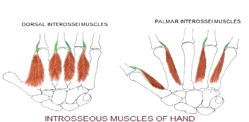
Intrinsic Hand Muscles. This image shows the dorsal and palmar interossei.
Contributed by Bordoni Bruno, PhD.
(Click Image to Enlarge)
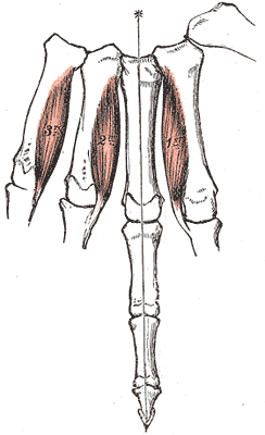
Palmar Interossei of the Hand. Volar view illustrating the palmar interossei muscles and their attachments from the metacarpal bones to the proximal phalanges and extensor hoods.
Henry Vandyke Carter, Public domain, via Wikimedia Commons
(Click Image to Enlarge)
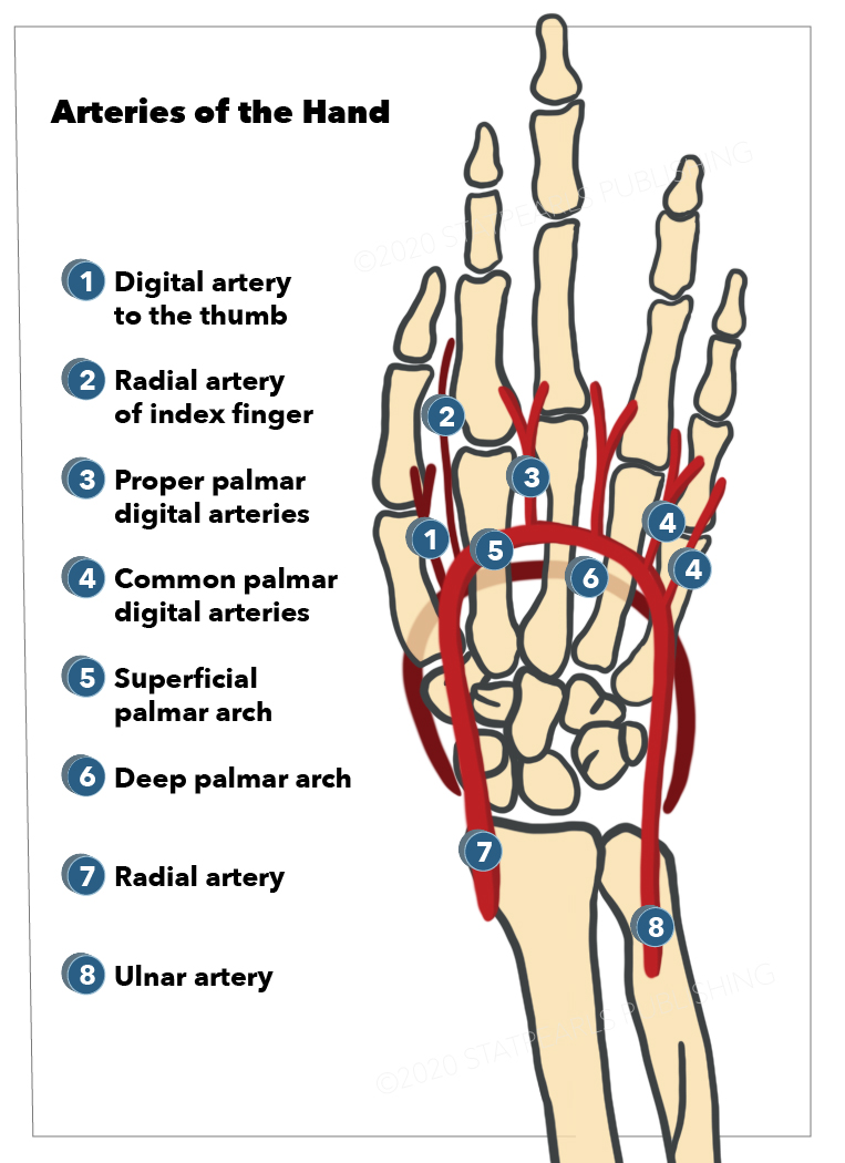
Arteries of the Hand. Shown here are the ulnar artery, radial artery, deep palmar arch, superficial palmar arch, common palmar digital arteries, proper palmar digital arteries, radial artery of the index finger (radialis indicis), and digital artery to the thumb (princeps pollicis).
StatPearls Publishing Illustration
(Click Image to Enlarge)
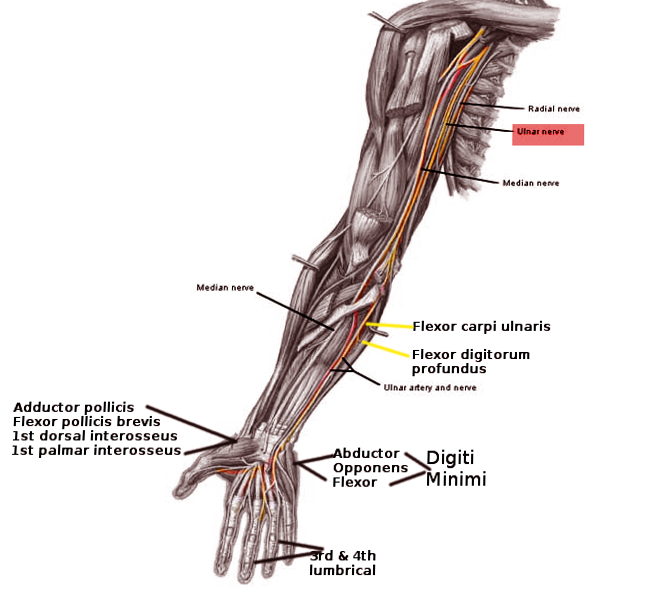
Ulnar Nerve Pathway. Anterior view illustrating the course of the ulnar nerve from the shoulder to the hand.
Image courtesy O.Chaigasame
References
Picasso R, Zaottini F, Pistoia F, Perez MM, Macciò M, Bianco D, Rinaldi S, Pansecchi M, Rossi G, Tovt L, Martinoli C. Ultrasound of the palmar aspect of the hand: normal anatomy and clinical applications of intrinsic muscles imaging. Journal of ultrasonography. 2023 Sep:23(94):e122-e130. doi: 10.15557/jou.2023.0021. Epub 2023 Sep 11 [PubMed PMID: 37732107]
Morrison PE, Hill RV. And then there were four: Anatomical observations on the pollical palmar interosseous muscle in humans. Clinical anatomy (New York, N.Y.). 2011 Nov:24(8):978-83. doi: 10.1002/ca.21253. Epub 2011 Aug 25 [PubMed PMID: 22009503]
Bello-Hellegouarch G, Aziz MA, Ferrero EM, Kern M, Francis N, Diogo R. "Pollical palmar interosseous muscle" (musculus adductor pollicis accessorius): attachments, innervation, variations, phylogeny, and implications for human evolution and medicine. Journal of morphology. 2013 Mar:274(3):275-93. doi: 10.1002/jmor.20090. Epub 2012 Oct 29 [PubMed PMID: 23109102]
Level 3 (low-level) evidenceTickle C. How the embryo makes a limb: determination, polarity and identity. Journal of anatomy. 2015 Oct:227(4):418-30. doi: 10.1111/joa.12361. Epub 2015 Aug 7 [PubMed PMID: 26249743]
Cole P, Kaufman Y, Hatef DA, Hollier LH Jr. Embryology of the hand and upper extremity. The Journal of craniofacial surgery. 2009 Jul:20(4):992-5. doi: 10.1097/SCS.0b013e3181abb18e. Epub [PubMed PMID: 19553860]
Wilde S, Feneck EM, Mohun TJ, Logan MPO. 4D formation of human embryonic forelimb musculature. Development (Cambridge, England). 2021 Feb 17:148(4):. doi: 10.1242/dev.194746. Epub 2021 Feb 17 [PubMed PMID: 33234713]
Poirot Y, Duparc F, Hue AG, Gandolfi S, Dacher JN, Auquit-Aukbur I. Cutaneous vascularization of the proximal two-thirds of the dorsal aspect of the hand: descriptive anatomical study of a perforating arterial arch. Surgical and radiologic anatomy : SRA. 2023 Sep:45(9):1073-1081. doi: 10.1007/s00276-023-03185-w. Epub 2023 Jul 12 [PubMed PMID: 37438569]
Granoff MD, Pardo JA, Johnson AR, Fleishman A, Tillotson E, Thomson S, Lee BT, Singhal D. Superficial and Functional Lymphatic Anatomy of the Upper Extremity. Plastic and reconstructive surgery. 2022 Oct 1:150(4):900-907. doi: 10.1097/PRS.0000000000009555. Epub 2022 Aug 4 [PubMed PMID: 35939638]
Yang T, Rui YJ. Innervation of the lumbrical and interosseous muscles in hand: analysis of distribution of nerve fascicles and quantification of their surface projections. Journal of plastic surgery and hand surgery. 2022 Oct:56(5):310-317. doi: 10.1080/2000656X.2021.1981348. Epub 2021 Sep 28 [PubMed PMID: 34581658]
Chambers SB, Wu KY, Smith C, Potra R, Ferreira LM, Gillis J. Interfascicular Anatomy of the Motor Branch of the Ulnar Nerve: A Cadaveric Study. The Journal of hand surgery. 2023 Mar:48(3):309.e1-309.e6. doi: 10.1016/j.jhsa.2021.10.012. Epub 2021 Dec 20 [PubMed PMID: 34949481]
Eladoumikdachi F, Valkov PL, Thomas J, Netscher DT. Anatomy of the intrinsic hand muscles revisited: part I. Interossei. Plastic and reconstructive surgery. 2002 Oct:110(5):1211-24 [PubMed PMID: 12360058]
Mejia A, Lichtig AE, Ghosh A, Balasubramaniyan A, Mass D, Amirouche F. The Effect of Metacarpal Shortening on Finger Strength and Joint Motion: A Cadaveric Biomechanical Study. Journal of hand surgery global online. 2023 Jul:5(4):407-412. doi: 10.1016/j.jhsg.2023.03.007. Epub 2023 Apr 6 [PubMed PMID: 37521540]
Chopra R, Hayton M, Dunbar PJ. Exercise induced chronic compartment syndrome of the first dorsal interosseous compartment of the hand: a case report. Hand (New York, N.Y.). 2009 Dec:4(4):415-7. doi: 10.1007/s11552-009-9203-x. Epub 2009 Jun 11 [PubMed PMID: 19517196]
Level 3 (low-level) evidenceAndrews K, Rowland A, Pranjal A, Ebraheim N. Cubital tunnel syndrome: Anatomy, clinical presentation, and management. Journal of orthopaedics. 2018 Sep:15(3):832-836. doi: 10.1016/j.jor.2018.08.010. Epub 2018 Aug 16 [PubMed PMID: 30140129]