Introduction
Recent advances in various surgical techniques, combined with the development of more minimally invasive procedures, have led to an increase in outpatient procedures. With these developments, analgesic techniques need to keep pace with these surgical advancements. Studies have shown that peripheral nerve blocks are usually well-tolerated and provide regional analgesia that is superior to other modalities, such as oral pain medications or general anesthesia.[1][2][3]
Anatomy and Physiology
Register For Free And Read The Full Article
 Search engine and full access to all medical articles
- 10 free questions in your specialty
- Free CME/CE Activities
- Free daily question in your email
- Save favorite articles to your dashboard
- Emails offering discounts
Learn more about a Subscription to StatPearls Point-of-Care
Anatomy and Physiology
Anatomy and landmarks depend on the type of block being performed. Please refer to the Technique or Treatment section for more information on the techniques of the more common peripheral nerve blocks.
Indications
No strict guidelines for the use of peripheral nerve blocks have been established. However, the general rationale is to implement regional blocks in cases where conservative measures have failed, or to avoid the adverse effects and complications associated with general anesthesia and oral medications. The following are examples of where peripheral nerve blocks may be preferable:
- Patients who are at high risk of respiratory depression related to general anesthesia
- Patients who want to avoid systemic medications
- Patients who are intolerant or not responsive to oral medications
- As adjunct therapy to reduce preoperative and postoperative opioid use [4]
Contraindications
Absolute contraindications to the use of peripheral nerve blocks include allergy to local anesthetics, inability to cooperate, or patient refusal. Experts advise postponing or reconsidering a nerve injection when an active infection at the injection site is present, preexisting neural deficits along with the distribution of the block are noted, or in patients with coagulopathies or on antithrombotic drugs.[5]
Equipment
The equipment used depends on the type of peripheral nerve block technique used, including:
- Nerve stimulator guidance: A peripheral nerve stimulator that delivers an adjustable electrical current to the tip of a hollow, insulated, disposable needle. The needle is attached to a syringe via specific tubing, allowing for the aspiration and injection of a local anesthetic. A wire connects the needle to an electrode, allowing the electrical pulse to be transmitted and stimulating the nerve.
- Ultrasound guidance: Portable ultrasound machines with both high and low-frequency probes that can identify superficial and deeper nerves [6]
- Continuous catheter: Numerous kits are available, typically containing a needle and catheter. A standard epidural kit can often be utilized.
Personnel
A well-versed medical professional, highly familiar and experienced with the type of peripheral nerve block being performed, should administer the specific injection.
Preparation
Taking a detailed medical history is essential to identify conditions, eg, coagulopathy or respiratory compromise, that may impact the decision to perform a peripheral nerve block. A thorough physical exam is also prudent to determine if any preexisting sensory or motor deficits are present in the distribution of the block. Studies show that patients with preexisting sensory or motor deficits may be more likely to develop new deficits following a block than patients without preexisting deficits. Following the history and physical examination, the patient should be informed about the risks, benefits, and care needed during the recovery phase of the block.
For patients who are receiving a peripheral nerve block for a surgical procedure, they should follow the same fasting guidelines for the surgery, as it may be necessary for deep sedation to be used in cases of an inadequate block. Also, intravenous access should be obtained due to the risk of potential complications like vasovagal events, local anesthetic toxicity, and the possible use of general anesthetics.
Technique or Treatment
Peripheral Nerve Block Types and Techniques
The technique for peripheral nerve blocks varies based on the type of block performed, including:
Interscalene block
An interscalene block anesthetizes nerve roots from the cervical plexus (C3, C4, supraclavicular nerve) and upper and middle trunks of the brachial plexus (C5-C7). For positioning, the patient is placed in a supine position with the head turned away from the side of the block. Sternal notch, the sternal and clavicular heads of the sternocleidomastoid muscle, and the clavicle are identified and marked. An ultrasound probe is placed in a transverse position with its long axis running across the neck, just above the clavicle. The carotid artery and internal jugular vein are visualized. The subclavian artery is identified by directing the beam towards the first rib. Nerves are then traced to cephalad. At the C6 nerves of the brachial plexus are visualized in a vertical orientation within the interscalene groove (see Image. Suprascapular Nerve Block). A needle is then placed in-plane or out-of-plane and directed toward the nerves. A needle tip is placed next to the nerve roots, and a total of 12 mL to 30 mL of local anesthetic is injected.
Supraclavicular block
The patient is placed in a supine position with their arms by their sides and their head turned away from the side where the block is being administered (see Image. Supraclavicular Ultrasound-Guided, Brachial Plexus Nerve Block). The ultrasound probe is placed in a transverse position just above the clavicle. The carotid artery and internal jugular vein are visualized, and the needle is inserted in-plane (parallel to the probe). A local anesthetic is injected to hydro-dissect between the nerves until the tip reaches an area bordered by the first rib, subclavian artery, and brachial plexus. Approximately 20 mL to 30 mL of local anesthetic is injected. Before injection, however, aspiration should be performed to ensure no blood return is noted.
Infraclavicular block
The patient is placed in the supine position with the head turned away from the side of the block. The patient's arm is abducted with the elbow flexed to identify the coracoid process. The axillary artery is identified, and the cords of the brachial plexus are visualized adjacent to the artery using ultrasound. The needle is placed adjacent to the axillary artery in the cranio-posterior quadrant, and 30 to 40 mL of local anesthetic is administered. Before injection, however, aspiration should be performed to ensure no blood returns.
Axillary block
This block anesthetizes the nerves of the brachial plexus at the level of individual nerves and often requires multiple injections. The patient is positioned supine with their arm abducted to 90 degrees and the elbow flexed at 90 degrees. The ultrasound transducer is placed transversely in the axilla, and the needle is introduced perpendicular to the skin and advanced until the tip is next to each nerve.
Intercostobrachial block
The patient is positioned supine with their arm abducted to expose the axillary fossa. The intercostobrachial nerve runs in the subcutaneous tissue of the medial upper arm. The needle is advanced subcutaneously across the medial aspect of the arm while injecting 5 to 10 mL of local anesthetic.
Radial nerve block
The radial nerve emerges between the brachioradialis tendon and the radius, just proximal to the styloid process. The needle is inserted subcutaneously, just proximal to the styloid process of the radius, aiming medially, and 3 to 5 mL of local anesthetic is injected.
Median nerve block
The median nerve block is located between the tendons of the flexor palmaris longus and the flexor carpi radialis. The needle is inserted between the two tendons until it penetrates the fascia and is advanced until contact is made with bone. The needle should be redirected and local anesthetic injected in lateral and medial directions (see Image. Median Nerve Block).
Ulnar nerve block
The ulnar nerve runs between the ulnar artery and the flexor carpi ulnaris tendon. The tendon is just superficial to the ulnar nerve. A needle is placed under the tendon close to its attachment just above the styloid process of the ulna and advanced 5 to 10 mm, and 3 to 5 mL of local anesthetic is injected at this location.
Lumbar plexus block
The patient is placed in the lateral decubitus position, with the operative side up, and the leg is flexed at the hip and knee. The ultrasound probe is placed longitudinally adjacent to the spine at the second to third lumbar level. The needle is inserted at the cephalad edge using the in-plane technique. The length of the needle should be seen as it approaches the target structure, which is the posterior third of the psoas major muscle.
Femoral nerve block
The patient is placed in a supine position. The femoral nerve is visualized using ultrasound imaging, located laterally to the artery. An in-plane or out-of-plane approach can be used, where the needle is inserted and the tip is placed adjacent to the nerve. Local anesthetic is then injected in 5 mL increments, ranging from 20 to 50 mL. Before injection, however, aspiration should be performed to ensure no blood return is noted.
Fascia iliaca block
The patient is placed in a supine position, where, using ultrasound, the probe is placed transversely to the leg at the junction of the middle and lateral thirds (between the ASIS and pubic tubercle) to identify the fascia lata, iliacus muscle, and fascia iliaca. The needle is introduced in-plane inferior to the inguinal ligament and guided beneath the fascia iliaca, and 30 mL of local anesthetic is injected in 5 mL increments. Before injection, however, aspiration should be performed to ensure no blood return is noted.[7]
Obturator nerve block
The patient is placed in the supine position with the leg externally rotated. The femoral vein is identified using an ultrasound probe placed in the inguinal crease. The probe is then moved medially to visualize the pectineus and adductor longus muscles. The needle is inserted in-plane or out of plane and is directed to the fascial plane between the adductor brevis and magnus, and 5 mL to 10 mL of local anesthetic is injected. Before injection, however, aspiration should be performed to ensure no blood return is noted.
Sciatic nerve block
A sciatic nerve block can be approached either anteriorly or posteriorly. For the posterior approach, the patient is placed in the lateral decubitus position, with the hip flexed to 45 degrees and the knee extended to 90 degrees. For the anterior approach, the patient is positioned in the same manner. The ultrasound probe is held transversely to the nerve's course, and the sciatic nerve is found lateral to the ischial tuberosity and deep to the gluteus maximus muscle. The needle is inserted in-plane from the lateral aspect of the transducer and positioned with the tip of the needle adjacent to the nerve. Approximately 20 mL of local anesthetic is injected in 5 mL increments with gentle aspirations between injections. Before injection, however, aspiration should be performed to ensure no blood return is noted.
Popliteal nerve block
The patient can be positioned in a prone, lateral decubitus, or supine position. Clinicians typically use 2 approaches. For the posterior approach, the biceps femoris and semitendinosus/semimembranosus tendons are palpated. The ultrasound probe is placed transversely to the thigh and in the popliteal crease. The popliteal artery is used as the landmark, and the tibial nerve is found superficial and lateral to the popliteal artery. The nerve is then followed cephalad to the point where the common fibular nerve joins the tibial nerve from the lateral side, forming the sciatic nerve. The sciatic nerve is blocked proximally to ensure that both the common fibular nerve and the tibial nerve are anesthetized.
Saphenous nerve block
For a saphenous nerve block, the patient is positioned supine with the leg straight. The saphenous nerve is identified as it exits from the adductor canal adjacent to the femoral artery using the ultrasound probe placed perpendicularly to the thigh at the midpoint between the anterior superior iliac spine and the distal end of the femur (see Image. Saphenous Nerve Block). As the nerve is followed distally, the saphenous nerve becomes more superficial, traveling with an arterial branch just deep to the sartorius muscle. Using an in-plane approach, 10 mL of local anesthetic is injected deep into the sartorius muscle at the lateral border of the artery.
Pericapsular nerve group block
The pericapsular nerve group (PENG) block is an interfascial plane block aiming to block articular branches supplied by femoral, obturator, and accessory obturator nerves. The PENG block is indicated for anterior hip arthroplasties, lateral hip arthroplasties, and hip fractures. This block is performed in supine position by depositing 15 to 20 mL of local anesthetic in the plane between the psoas tendon and the pubic ramus under direct ultrasound visualization. Femoral nerve blocks, fascia iliaca compartment blocks, or lumbar plexus blocks have also been used to manage postoperative analgesia in hip surgeries. However, these blocks result in weakness of the quadriceps muscles and thus predispose to falls. These blocks also result in incomplete analgesia of the hip, as they spare a few articular branches to the hip. Conversely, the main advantage of the PENG block is that it provides better analgesia of the hip without causing any muscle weakness. Since no muscle weakness occurs, the patient can participate in physical therapy early.
IPACK block
An IPACK block is an acronym for infiltration of local anesthetic into the interspace between the popliteal artery and the posterior capsule of the knee. This technique was first introduced by Dr. Sanjay K Sinha at the American Society of Regional Anesthesia (ASRA) meeting in 2012.
The IPACK block is used for postoperative analgesia for total knee arthroplasty and cruciate ligament repair. Posterior knee pain is mediated by articular branches arising mainly from the tibial nerve, with contributions from the obturator nerve. In the IPACK block, 15 to 20 mL of local anesthetic is deposited under ultrasound guidance in the tissue plane of the femoral artery and the posterior aspect of the capsule of the knee joint. The main advantage of the IPACK block is that it is a muscle-sparing block, which does not result in foot drop or loss of sensorimotor function in the leg and foot.
Complications
Potential complications and adverse effects depend on the type of block performed. However, complications include peripheral nerve injury (although not common, the rate may be as high as 8% to 10%), hematoma, local anesthetic systemic toxicity, allergic reaction, infection, and a secondary injury, which includes reduced sensation after nerve block (ie, post-block neurological dysfunction).[8][9][10][11]
Clinical Significance
Peripheral nerve blocks hold significant clinical importance as they offer targeted regional anesthesia and analgesia, particularly crucial in the context of modern surgical advancements and the increasing trend toward outpatient procedures. As minimally invasive surgeries become more common, a growing need for analgesic techniques that match the precision and efficiency of these surgical methods has been demonstrated. Peripheral nerve blocks provide superior pain control compared to systemic opioids or general anesthesia, reducing the risk of systemic adverse effects such as respiratory depression, nausea, sedation, and opioid dependence.[12] They are especially beneficial for patients who are at high risk for complications from general anesthesia, intolerant to oral pain medications, or seeking to minimize opioid use postoperatively. In many cases, peripheral nerve blocks enable faster recovery, earlier mobilization, and improved participation in physical therapy, which is crucial for optimizing surgical outcomes.
Clinically, peripheral nerve blocks are versatile and can be tailored to the specific anatomical region and surgical procedure, from interscalene and supraclavicular blocks for upper limb surgeries to femoral and sciatic nerve blocks for lower extremity operations. Specialized blocks, such as the PENG block for hip surgeries and the IPACK block for knee procedures, highlight the evolution of nerve blocks toward techniques that offer excellent pain relief while preserving muscle strength and minimizing functional impairment. This muscle-sparing advantage enhances postoperative rehabilitation and reduces the risk of falls, especially in elderly populations. Although generally safe, clinicians must remain vigilant for potential complications, including nerve injury, hematoma, systemic toxicity, infection, and post-block neurological dysfunction. Appropriate patient selection, strict adherence to aseptic technique, and careful ultrasound-guided administration are essential to maximize benefits and minimize risks. Overall, peripheral nerve blocks represent a critical component of modern perioperative pain management, aligning with the broader goals of enhanced recovery protocols and improved patient satisfaction.
Enhancing Healthcare Team Outcomes
Effective implementation of peripheral nerve blocks requires an interprofessional approach that prioritizes patient-centered care, safety, and optimal clinical outcomes. Physicians—including anesthesiologists, surgeons, and emergency medicine clinicians—play a central role in determining the appropriate block type, performing the procedure under ultrasound guidance, and assessing individual risk factors such as coagulation status or local anesthetic allergies. Their responsibilities also include ensuring informed consent, accurate documentation, and the availability of resuscitation equipment before initiation. Advanced practitioners, such as nurse anesthetists or physician assistants, contribute by supporting block placement, monitoring for complications, and reinforcing perioperative education with the patient. Skilled ultrasound use and proper needle placement techniques are critical competencies to minimize risks such as nerve injury, hematoma, or systemic toxicity. A standardized protocol should guide every aspect of the block, from preparation and patient positioning to post-procedural observation, to promote consistency, safety, and effectiveness.
Interprofessional communication and care coordination are vital to the success of peripheral nerve blocks. Nurses are essential team members responsible for real-time monitoring of the patient’s vital signs, providing preprocedural and postprocedural care, and promptly reporting signs of complications. Pharmacists enhance safety by verifying the correct local anesthetic dose, screening for drug interactions, and preparing medications as needed. Collaboration among all healthcare professionals ensures rapid response to adverse events and supports adherence to safety protocols. Clear communication, shared decision-making, and defined roles within the care team help streamline workflow and reduce variability in practice. Together, these strategies enhance team performance, reduce opioid reliance, improve pain control, and ultimately lead to better patient satisfaction and recovery outcomes in the context of peripheral nerve block use.
Media
(Click Image to Enlarge)
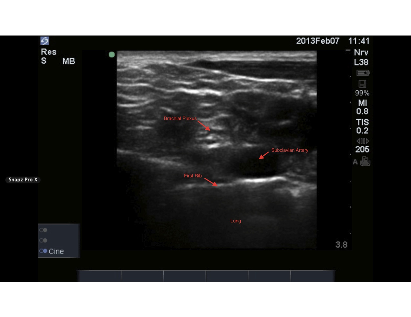
Supraclavicular Ultrasound-Guided, Brachial Plexus Nerve Block. A local anesthetic is injected to hydro-dissect between the nerves until the tip reaches an area bordered by the first rib, subclavian artery, and brachial plexus.
StatPearls
(Click Video to Play)
Median Nerve Block. Live image of ultrasound-guided plane median nerve block, flexor digitorum superficialis, flexor digitorum profundus.
Contributed by J Pester, DO
(Click Image to Enlarge)
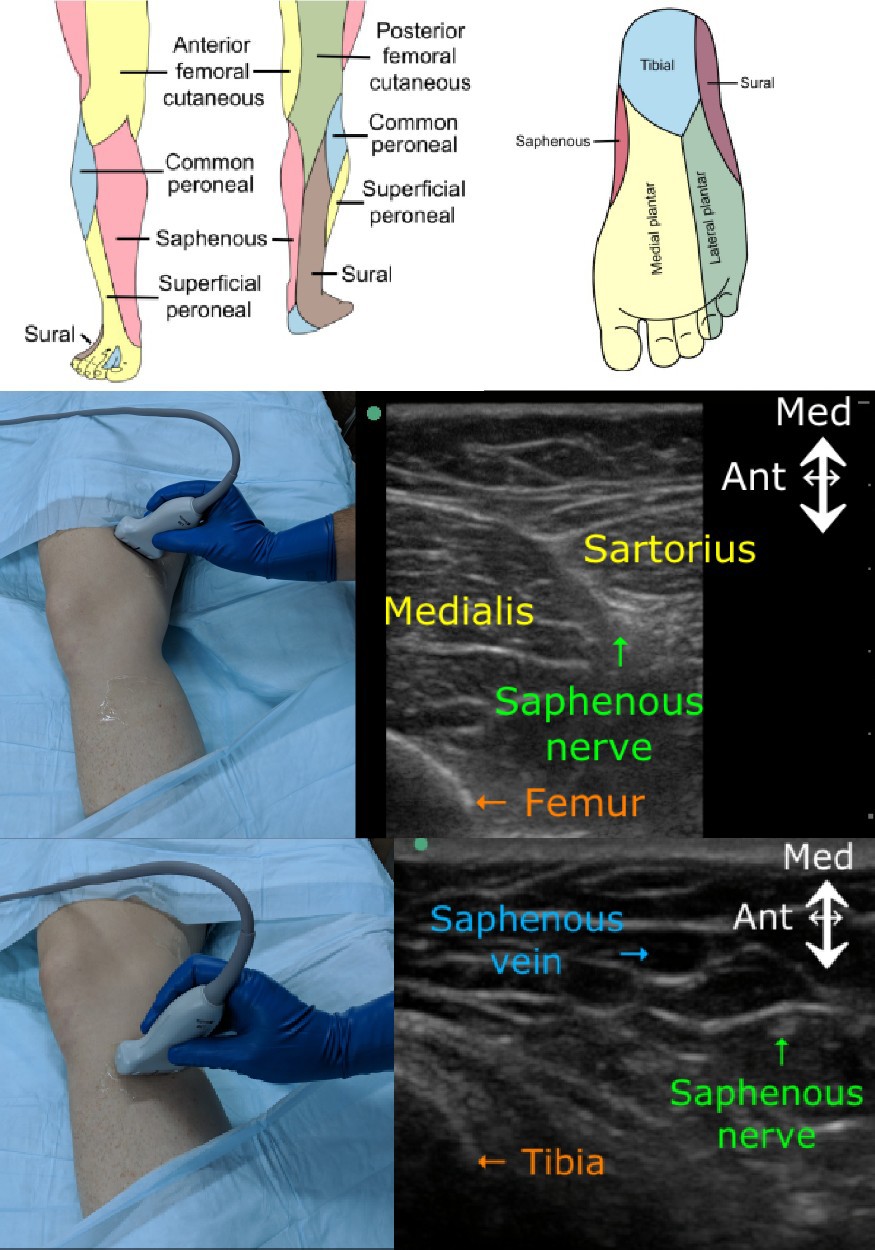
Saphenous Nerve Block. Image of saphenous nerve block.
Contributed by M Brady, MD
(Click Video to Play)
Suprascapular Nerve Block. Video demonstrates ultrasound-guided suprascapular nerve block at the level of the suprascapular notch.
Contributed by E Helm, MD
References
Kurita GP, Sjøgren P, Klepstad P, Mercadante S. Interventional Techniques to Management of Cancer-Related Pain: Clinical and Critical Aspects. Cancers. 2019 Mar 29:11(4):. doi: 10.3390/cancers11040443. Epub 2019 Mar 29 [PubMed PMID: 30934870]
Raj N. Regional anesthesia for sternotomy and bypass-Beyond the epidural. Paediatric anaesthesia. 2019 May:29(5):519-529. doi: 10.1111/pan.13626. Epub [PubMed PMID: 30861264]
Donado C, Solodiuk J, Rangel SJ, Nelson CP, Heeney MM, Mahan ST, Ullrich C, Tsegaye B, Berde CB. Patient- and Nurse-Controlled Analgesia: 22-Year Experience in a Pediatric Hospital. Hospital pediatrics. 2019 Feb:9(2):129-133. doi: 10.1542/hpeds.2018-0179. Epub 2019 Jan 17 [PubMed PMID: 30655310]
Guay J, Kopp S. Peripheral nerve blocks for hip fractures in adults. The Cochrane database of systematic reviews. 2020 Nov 25:11(11):CD001159. doi: 10.1002/14651858.CD001159.pub3. Epub 2020 Nov 25 [PubMed PMID: 33238043]
Level 1 (high-level) evidenceEcoffey C,Bosenberg A,Lonnqvist PA,Suresh S,Delbos A,Ivani G, Practice advisory on the prevention and management of complications of pediatric regional anesthesia. Journal of clinical anesthesia. 2022 Aug [PubMed PMID: 35313269]
Saranteas T, Kostroglou A, Efstathiou G, Giannoulis D, Moschovaki N, Mavrogenis AF, Perisanidis C. Peripheral nerve blocks in the cervical region: from anatomy to ultrasound-guided techniques. Dento maxillo facial radiology. 2020 Dec 1:49(8):20190400. doi: 10.1259/dmfr.20190400. Epub 2020 Mar 16 [PubMed PMID: 32176537]
Simić A, Nesek Adam V, Rošić D, Kočet N, Svetec M, Herceg A, Keranović A, Rašić Ž. PERIPHERAL NERVE BLOCKS FOR HIP FRACTURES IN EMERGENCY MEDICINE. Acta clinica Croatica. 2022 Jun:61(Suppl 1):78-83. doi: 10.20471/acc.2022.61.s1.13. Epub [PubMed PMID: 36304813]
Lemke E, Johnston DF, Behrens MB, Seering MS, McConnell BM, Swaran Singh TS, Sondekoppam RV. Neurological injury following peripheral nerve blocks: a narrative review of estimates of risks and the influence of ultrasound guidance. Regional anesthesia and pain medicine. 2024 Feb 5:49(2):122-132. doi: 10.1136/rapm-2023-104855. Epub 2024 Feb 5 [PubMed PMID: 37940348]
Level 3 (low-level) evidenceSaranteas T, Koliantzaki I, Savvidou O, Tsoumpa M, Eustathiou G, Kontogeorgakos V, Souvatzoglou R. Acute pain management in trauma: anatomy, ultrasound-guided peripheral nerve blocks and special considerations. Minerva anestesiologica. 2019 Jul:85(7):763-773. doi: 10.23736/S0375-9393.19.13145-8. Epub 2019 Feb 7 [PubMed PMID: 30735016]
Tran DQ, Salinas FV, Benzon HT, Neal JM. Lower extremity regional anesthesia: essentials of our current understanding. Regional anesthesia and pain medicine. 2019 Jan 11:():. pii: rapm-2018-000019. doi: 10.1136/rapm-2018-000019. Epub 2019 Jan 11 [PubMed PMID: 30635506]
Level 3 (low-level) evidenceHussain N, McCartney CJL, Neal JM, Chippor J, Banfield L, Abdallah FW. Local anaesthetic-induced myotoxicity in regional anaesthesia: a systematic review and empirical analysis. British journal of anaesthesia. 2018 Oct:121(4):822-841. doi: 10.1016/j.bja.2018.05.076. Epub 2018 Aug 8 [PubMed PMID: 30236244]
Level 1 (high-level) evidenceAnger M, Valovska T, Beloeil H, Lirk P, Joshi GP, Van de Velde M, Raeder J, PROSPECT Working Group* and the European Society of Regional Anaesthesia and Pain Therapy. PROSPECT guideline for total hip arthroplasty: a systematic review and procedure-specific postoperative pain management recommendations. Anaesthesia. 2021 Aug:76(8):1082-1097. doi: 10.1111/anae.15498. Epub 2021 May 20 [PubMed PMID: 34015859]
Level 1 (high-level) evidence